Microscopy with undetected photons in the mid-infrared
Publication
Metrics
Paper Preview
Abstract
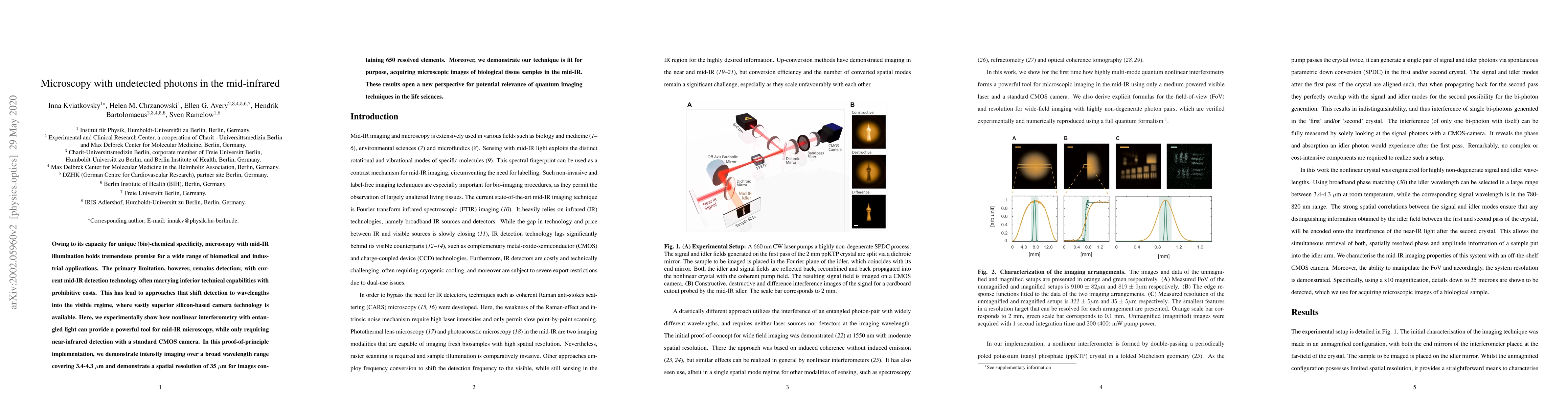
Owing to its capacity for unique (bio)-chemical specificity, microscopy withmid-IR illumination holds tremendous promise for a wide range of biomedical and industrial applications. The primary limitation, however, remains detection; with current mid-IR detection technology often marrying inferior technical capabilities with prohibitive costs. This has lead to approaches that shift detection towavelengths into the visible regime, where vastly superior silicon-based cameratechnology is available. Here, we experimentally show how nonlinear interferometry with entangled light can provide a powerful tool for mid-IR microscopy, while only requiring near-infrared detection with a standard CMOS camera. In this proof-of-principle implementation, we demonstrate intensity imaging overa broad wavelength range covering 3.4-4.3um and demonstrate a spatial resolution of 35um for images containing 650 resolved elements. Moreover, we demonstrate our technique is fit for purpose, acquiring microscopic images of biological tissue samples in the mid-IR. These results open a new perspective for potential relevance of quantum imaging techniques in the life sciences.
AI Key Findings
Get AI-generated insights about this paper's methodology, results, significance, and more — seven facets brought into focus.
Impact
Paper Details
Authors
PDF Preview
Key Terms
Citation Network
Current paper (gray), citations (green), references (blue)
Display is limited for performance on very large graphs.
Discussion 0