Microstructure.jl: a Julia Package for Probabilistic Microstructure Model Fitting with Diffusion MRI
Publication
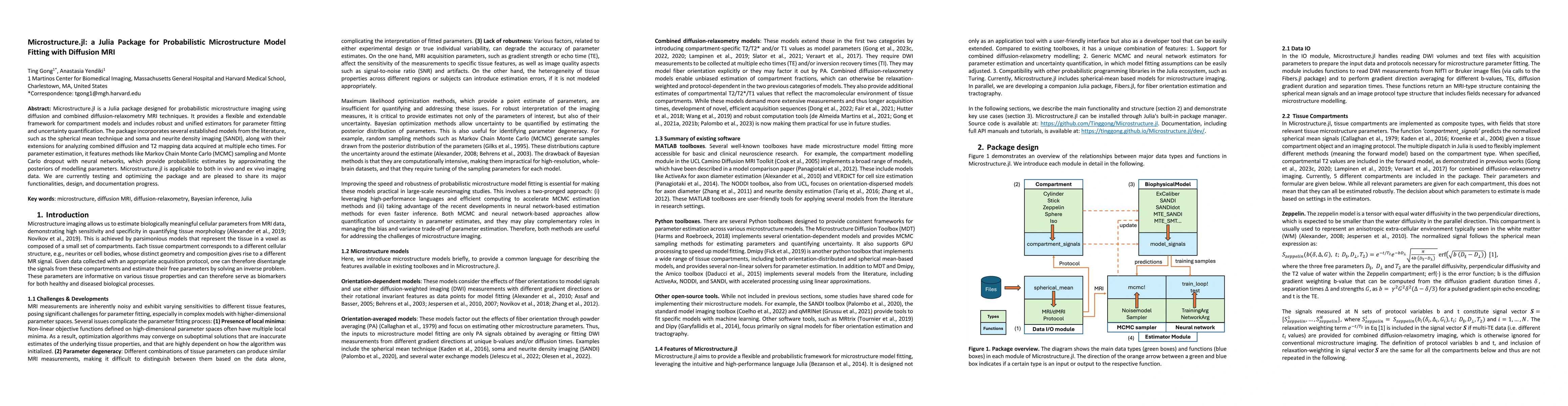
Metrics
AI Quick Summary
Microstructure.jl is a Julia package for probabilistic imaging using diffusion MRI, offering flexible frameworks for compartment models and robust estimators for parameter fitting. It supports various models and methods like MCMC for parameter estimation, applicable to both in vivo and ex vivo data.
Paper Preview
Abstract
Microstructure.jl is a Julia package designed for probabilistic microstructure imaging using diffusion and combined diffusion-relaxometry MRI techniques. It provides a flexible and extendable framework for compartment models and includes robust and unified estimators for parameter fitting and uncertainty quantification. The package incorporates several established models from the literature, such as the spherical mean technique and soma and neurite density imaging (SANDI), along with their extensions for analyzing combined diffusion and T2 mapping data acquired at multiple echo times. For parameter estimation, it features methods like Markov Chain Monte Carlo (MCMC) sampling and Monte Carlo dropout with neural networks, which provide probabilistic estimates by approximating the posteriors of modelling parameters. Microstructure.jl is applicable to both in vivo and ex vivo imaging data. We are currently testing and optimizing the package and are pleased to share its major functionalities, design, and documentation progress.
AI Key Findings
Get AI-generated insights about this paper's methodology, results, significance, and more — seven facets brought into focus.
Impact
Paper Details
Authors
PDF Preview
Citation Network
Current paper (gray), citations (green), references (blue)
Display is limited for performance on very large graphs.
Discussion 0