Microtubule Tracking in Electron Microscopy Volumes
Publication
Metrics
AI Quick Summary
Researchers developed a new method for tracking microtubules in electron microscopy volumes, achieving significant speed-ups and accuracy gains compared to prior methods.
Paper Preview
Abstract
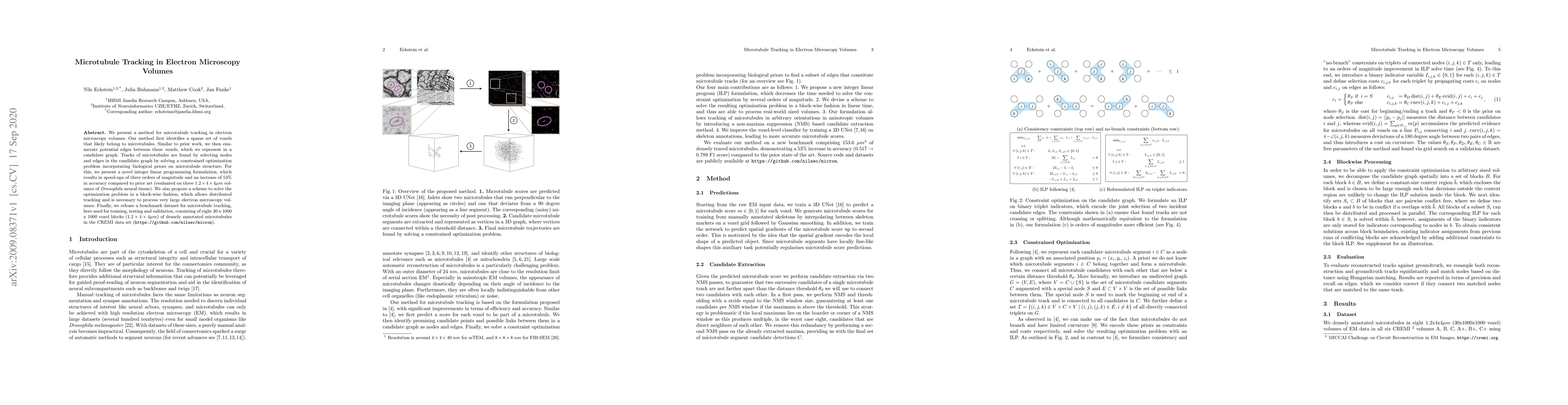
We present a method for microtubule tracking in electron microscopy volumes. Our method first identifies a sparse set of voxels that likely belong to microtubules. Similar to prior work, we then enumerate potential edges between these voxels, which we represent in a candidate graph. Tracks of microtubules are found by selecting nodes and edges in the candidate graph by solving a constrained optimization problem incorporating biological priors on microtubule structure. For this, we present a novel integer linear programming formulation, which results in speed-ups of three orders of magnitude and an increase of 53% in accuracy compared to prior art (evaluated on three 1.2 x 4 x 4$\mu$m volumes of Drosophila neural tissue). We also propose a scheme to solve the optimization problem in a block-wise fashion, which allows distributed tracking and is necessary to process very large electron microscopy volumes. Finally, we release a benchmark dataset for microtubule tracking, here used for training, testing and validation, consisting of eight 30 x 1000 x 1000 voxel blocks (1.2 x 4 x 4$\mu$m) of densely annotated microtubules in the CREMI data set (https://github.com/nilsec/micron).
AI Key Findings
Get AI-generated insights about this paper's methodology, results, significance, and more — seven facets brought into focus.
Impact
Paper Details
Authors
PDF Preview
Key Terms
Citation Network
Current paper (gray), citations (green), references (blue)
Display is limited for performance on very large graphs.
Discussion 0