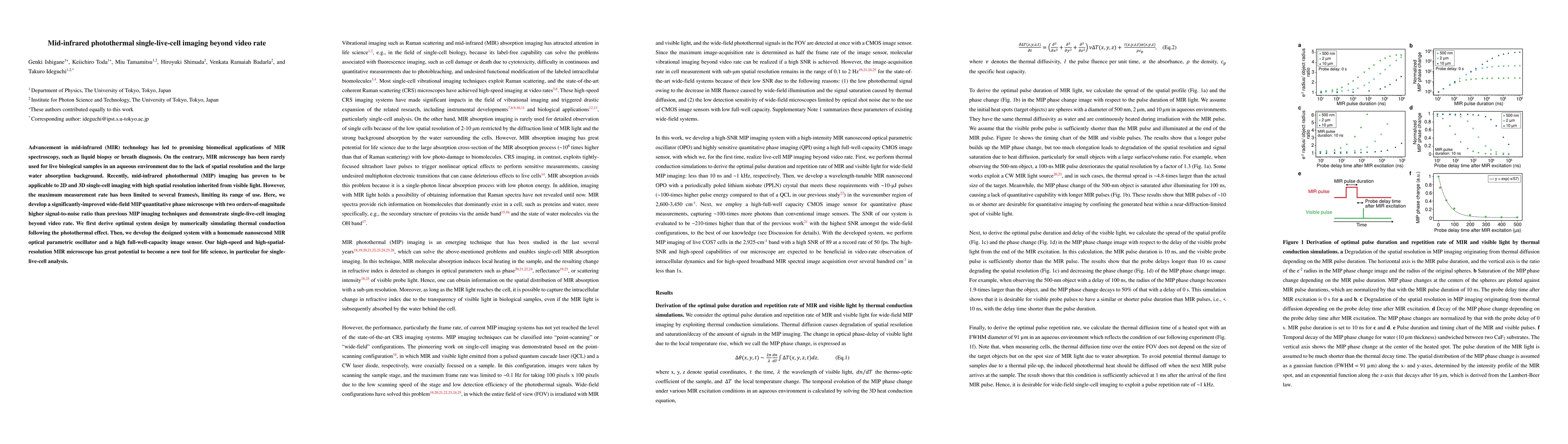
Mid-infrared photothermal single-live-cell imaging beyond video rate
Publication
Metrics
AI Quick Summary
This paper develops a high-speed, high-resolution mid-infrared photothermal microscope capable of imaging single live cells beyond video rate, overcoming previous limitations in spatial resolution and signal-to-noise ratio for aqueous biological samples. The improved system design and novel optical components promise new applications in life sciences.
Paper Preview
Abstract
Advancement in mid-infrared (MIR) technology has led to promising biomedical applications of MIR spectroscopy, such as liquid biopsy or breath diagnosis. On the contrary, MIR microscopy has been rarely used for live biological samples in an aqueous environment due to the lack of spatial resolution and the large water absorption background. Recently, mid-infrared photothermal (MIP) imaging has proven to be applicable to 2D and 3D single-cell imaging with high spatial resolution inherited from visible light. However, the maximum measurement rate has been limited to several frames/s, limiting its range of use. Here, we develop a significantly-improved wide-field MIP quantitative phase microscope with two orders-of-magnitude higher signal-to-noise ratio than previous MIP imaging techniques and demonstrate single-live-cell imaging beyond video rate. We first derive optimal system design by numerically simulating thermal conduction following the photothermal effect. Then, we develop the designed system with a homemade nanosecond MIR optical parametric oscillator and a high full-well-capacity image sensor. Our high-speed and high-spatial-resolution MIR microscope has great potential to become a new tool for life science, in particular for single-live-cell analysis.
AI Key Findings
Get AI-generated insights about this paper's methodology, results, significance, and more — seven facets brought into focus.
Impact
Paper Details
Authors
PDF Preview
Key Terms
Citation Network
Current paper (gray), citations (green), references (blue)
Display is limited for performance on very large graphs.
Discussion 0