Publication
Metrics
AI Quick Summary
MINFLUX is a groundbreaking super-resolution microscopy technique achieving nanometer resolution by scanning a patterned excitation beam to precisely localize single fluorophores, significantly outperforming camera-based methods in spatial and temporal resolution. Its high photon efficiency and potential for long-term tracking highlight its promise for advanced structural biology applications.
Paper Preview
Abstract
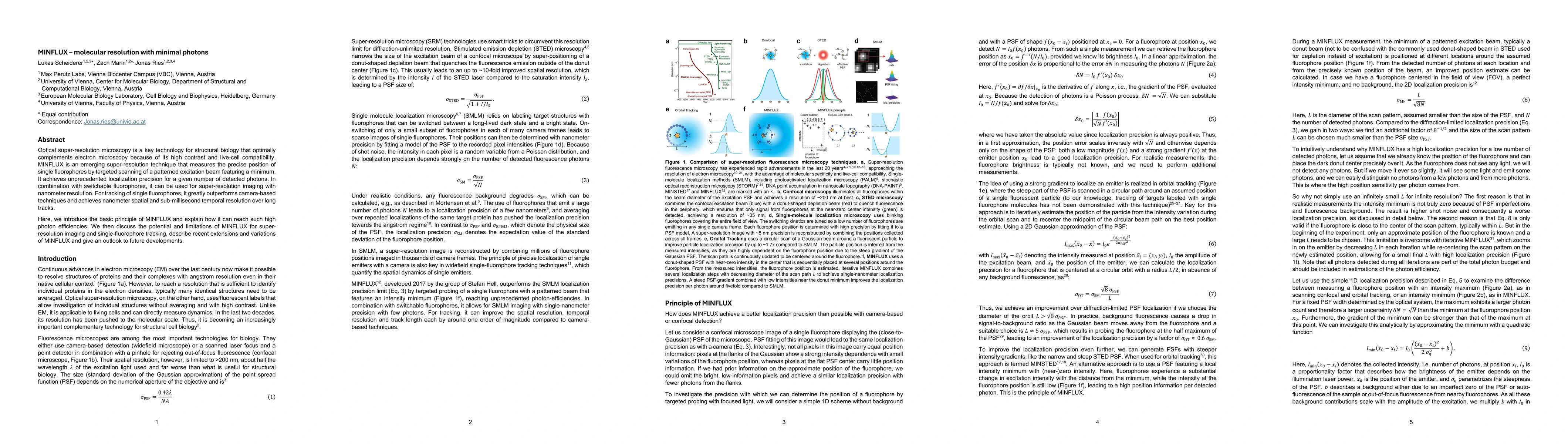
Optical super-resolution microscopy is a key technology for structural biology that optimally complements electron microscopy because of its high contrast and live-cell compatibility. MINFLUX is an emerging super-resolution technique that measures the precise position of single fluorophores by targeted scanning of a patterned excitation beam featuring a minimum. It achieves unprecedented localization precision for a given number of detected photons. In combination with switchable fluorophores, it can be used for super-resolution imaging with nanometer resolution. For tracking of single fluorophores, it greatly outperforms camera-based techniques and achieves nanometer spatial and sub-millisecond temporal resolution over long tracks. Here, we introduce the basic principle of MINFLUX and explain how it can reach such high photon efficiencies. We then discuss the potential and limitations of MINFLUX for super-resolution imaging and single-fluorophore tracking, describe recent extensions and variations of MINFLUX and give an outlook to future developments.
AI Key Findings
Get AI-generated insights about this paper's methodology, results, significance, and more — seven facets brought into focus.
Impact
Paper Details
Authors
PDF Preview
Citation Network
Current paper (gray), citations (green), references (blue)
Display is limited for performance on very large graphs.
Discussion 0