Summary
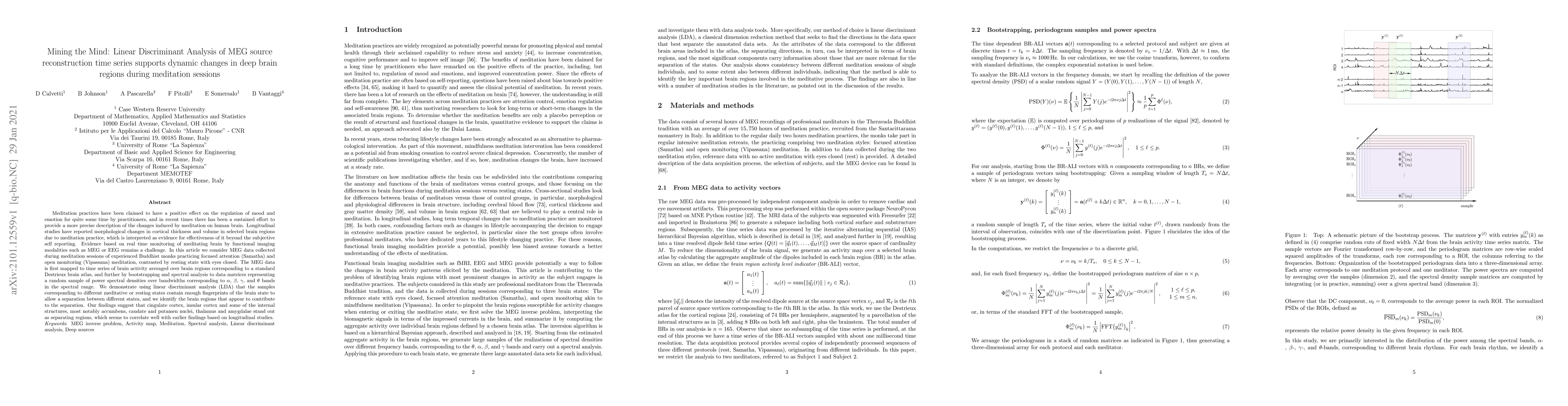
Meditation practices have been claimed to have a positive effect on the regulation of mood and emotion for quite some time by practitioners, and in recent times there has been a sustained effort to provide a more precise description of the changes induced by meditation on human brain. Longitudinal studies have reported morphological changes in cortical thickness and volume in selected brain regions due to meditation practice, which is interpreted as evidence for effectiveness of it beyond the subjective self reporting. Evidence based on real time monitoring of meditating brain by functional imaging modalities such as MEG or EEG remains a challenge. In this article we consider MEG data collected during meditation sessions of experienced Buddhist monks practicing focused attention (Samatha) and open monitoring (Vipassana) meditation, contrasted by resting state with eyes closed. The MEG data is first mapped to time series of brain activity averaged over brain regions corresponding to a standard Destrieux brain atlas, and further by bootstrapping and spectral analysis to data matrices representing a random sample of power spectral densities over bandwidths corresponding to $\alpha$, $\beta$, $\gamma$, and $\theta$ bands in the spectral range. We demonstrate using linear discriminant analysis (LDA) that the samples corresponding to different meditative or resting states contain enough fingerprints of the brain state to allow a separation between different states, and we identify the brain regions that appear to contribute to the separation. Our findings suggest that cingulate cortex, insular cortex and some of the internal structures, most notably accumbens, caudate and putamen nuclei, thalamus and amygdalae stand out as separating regions, which seems to correlate well with earlier findings based on longitudinal studies.
AI Key Findings
Get AI-generated insights about this paper's methodology, results, and significance.
Paper Details
PDF Preview
Key Terms
Citation Network
Current paper (gray), citations (green), references (blue)
Display is limited for performance on very large graphs.
Similar Papers
Found 4 papers| Title | Authors | Year | Actions |
|---|
Comments (0)