PET/CT imaging is the gold standard for tumor detection, offering high
accuracy in identifying local and metastatic lesions. Radiologists often begin
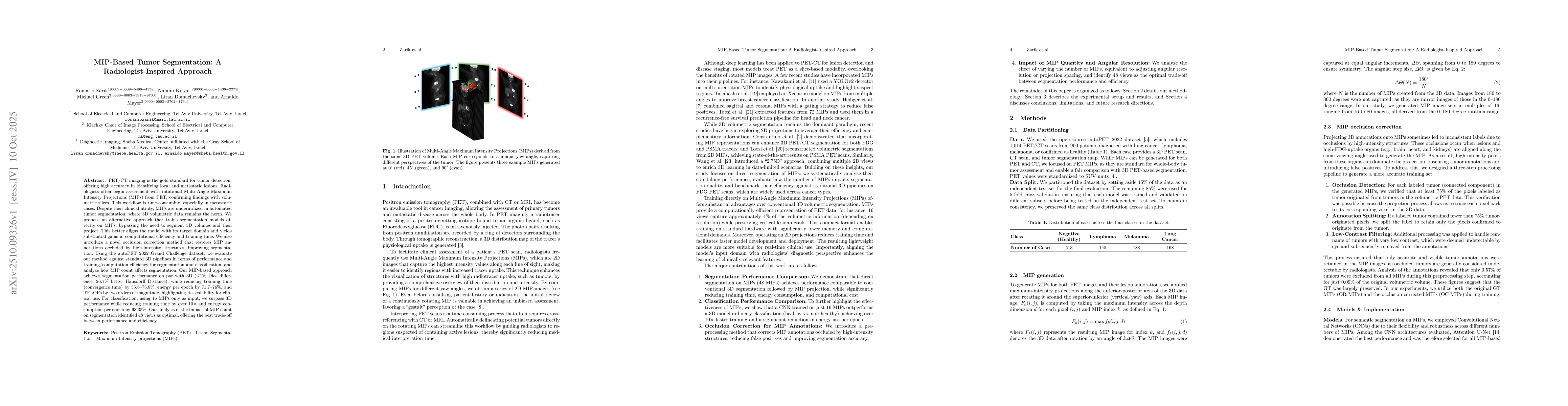
assessment with rotational Multi-Angle Maximum Intensity Projections (MIPs)
from PET, confirming findings with volumetric slices. This workflow is
time-consuming, especially in metastatic cases. Despite their clinical utility,
MIPs are underutilized in automated tumor segmentation, where 3D volumetric
data remains the norm. We propose an alternative approach that trains
segmentation models directly on MIPs, bypassing the need to segment 3D volumes
and then project. This better aligns the model with its target domain and
yields substantial gains in computational efficiency and training time. We also
introduce a novel occlusion correction method that restores MIP annotations
occluded by high-intensity structures, improving segmentation. Using the
autoPET 2022 Grand Challenge dataset, we evaluate our method against standard
3D pipelines in terms of performance and training/computation efficiency for
segmentation and classification, and analyze how MIP count affects
segmentation. Our MIP-based approach achieves segmentation performance on par
with 3D (<=1% Dice difference, 26.7% better Hausdorff Distance), while reducing
training time (convergence time) by 55.8-75.8%, energy per epoch by 71.7-76%,
and TFLOPs by two orders of magnitude, highlighting its scalability for
clinical use. For classification, using 16 MIPs only as input, we surpass 3D
performance while reducing training time by over 10x and energy consumption per
epoch by 93.35%. Our analysis of the impact of MIP count on segmentation
identified 48 views as optimal, offering the best trade-off between performance
and efficiency.
Discussion 0