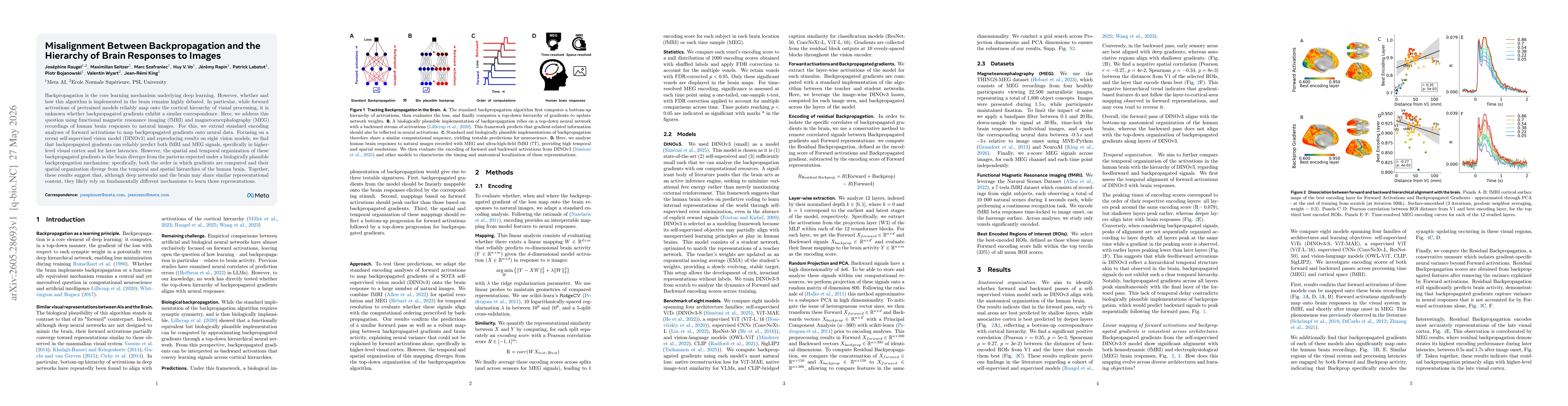
The study uses an encoding analysis framework to map both forward activations and backpropagated gradients from a diverse set of vision models (notably DINOv3) onto human brain data. It extends standard forward-activation encoding to include backpropagated gradients, applying linear Ridge regression with regularization to predict fMRI and MEG/EEG responses to natural images. The approach leverages high-spatial-resolution 7T fMRI and high-temporal-resolution MEG data, using cross-validation across stimuli to assess alignment, and compares representations across multiple models and layers to determine hierarchical correspondence and divergence from biological backpropagation.
Misalignment Between Backpropagation and the Hierarchy of Brain Responses to Images
Publication
Metrics
AI Quick Summary
The study shows that while gradient signals from deep learning models can predict human brain responses to images, their brain-like organization differs from how the brain processes information over time and space. This suggests that brains and deep networks may learn similar concepts but use fundamentally different learning mechanisms.
Quick Answers
What is "Misalignment Between Backpropagation and the Hierarchy of Brain Responses to Images" about?
The study shows that while gradient signals from deep learning models can predict human brain responses to images, their brain-like organization differs from how the brain processes information over time and space. This suggests that brains and deep networks may learn similar concepts but use fundamentally different learning mechanisms.
What methodology did the authors use?
The study uses an encoding analysis framework to map both forward activations and backpropagated gradients from a diverse set of vision models (notably DINOv3) onto human brain data. It extends standard forward-activation encoding to include backpropagated gradients, applying linear Ridge regression with regularization to predict fMRI and MEG/EEG responses to natural images. The approach leverages high-spatial-resolution 7T fMRI and high-temporal-resolution... More in Methodology →
What are the key results?
Backpropagated gradients can reliably predict both fMRI and MEG signals, especially in higher-level visual cortex and at later latencies, indicating a correspondence between model gradients and brain responses beyond forward activations. — The spatial and temporal organization of backpropagated gradients in the brain diverges from the biologically plausible backpropagation pattern: the order of gradient computations and their spatial arrangement do... More in Key Results →
Why is this work significant?
Demonstrates that while deep networks and the human visual system share representational content, the mechanisms by which they learn and organize information may be fundamentally different; backpropagation-like signals can map onto brain activity but do not mirror the brain's hierarchical timing and spatial structure, challenging assumptions about neural implementations of backpropagation. More in Significance →
What are the main limitations?
Study focuses on a subset of models (notably DINOv3) and a limited set of natural images, which may constrain generalizability across architectures and stimuli. — Inference relies on linear encoders; non-linear or more complex mappings between model gradients and brain responses could reveal different patterns. More in Limitations →
Abstract
Backpropagation is the core learning mechanism underlying deep learning. However, whether and how this algorithm is implemented in the brain remains highly debated. In particular, while forward activations of pretrained models reliably map onto the cortical hierarchy of visual processing, it is unknown whether backpropagated gradients exhibit a similar correspondence. Here, we address this question using functional magnetic resonance imaging (fMRI) and magnetoencephalography (MEG) recordings of human brain responses to natural images. For this, we extend standard encoding analyses of forward activations to map backpropagated gradients onto neural data. Focusing on a recent self-supervised vision model (DINOv3) and reproducing results on eight vision models, we find that backpropagated gradients can reliably predict both fMRI and MEG signals, specifically in higher-level visual cortex and for later latencies. However, the spatial and temporal organization of these backpropagated gradients in the brain diverges from the patterns expected under a biologically plausible backpropagation mechanism: specifically, both the order in which gradients are computed and their spatial organization diverge from the temporal and spatial hierarchies of the human brain. Together, these results suggest that, although deep networks and the brain may share similar representational content, they likely rely on fundamentally different mechanisms to learn those representations.
Key Findings, in focus
Seven facets of this paper, analysed and brought into focus by AI.
Demonstrates that while deep networks and the human visual system share representational content, the mechanisms by which they learn and organize information may be fundamentally different; backpropagation-like signals can map onto brain activity but do not mirror the brain's hierarchical timing and spatial structure, challenging assumptions about neural implementations of backpropagation.
- Backpropagated gradients can reliably predict both fMRI and MEG signals, especially in higher-level visual cortex and at later latencies, indicating a correspondence between model gradients and brain responses beyond forward activations.
- The spatial and temporal organization of backpropagated gradients in the brain diverges from the biologically plausible backpropagation pattern: the order of gradient computations and their spatial arrangement do not match the brain's hierarchical processing order.
- Forward activations align with the cortical hierarchy as expected, but the backpropagated gradient mappings reveal a mismatch in how learning signals would propagate top-down in the brain.
Demonstrates that while deep networks and the human visual system share representational content, the mechanisms by which they learn and organize information may be fundamentally different; backpropagation-like signals can map onto brain activity but do not mirror the brain's hierarchical timing and spatial structure, challenging assumptions about neural implementations of backpropagation.
Extends standard encoding analyses to map backpropagated gradients from state-of-the-art self-supervised vision models onto human brain data (fMRI and MEG), providing the first direct investigation of top-down gradient correspondence with neural responses and revealing divergence in spatial-temporal organization from biological backpropagation.
Pioneering direct test of whether backpropagated gradients align with brain activity across cortical hierarchies, showing reliable brain predictivity but non-biological top-down organization, thereby distinguishing representational similarity from learning mechanism similarity.
- Study focuses on a subset of models (notably DINOv3) and a limited set of natural images, which may constrain generalizability across architectures and stimuli.
- Inference relies on linear encoders; non-linear or more complex mappings between model gradients and brain responses could reveal different patterns.
- MEG and fMRI have different sensitivity profiles; integration assumptions may influence the interpretation of temporal vs. spatial alignment.
- Explore a broader range of architectures and training regimes to assess robustness of gradient-brain mappings.
- Investigate non-linear encoding models or kernel methods to capture subtler correspondences between gradients and neural data.
- Examine developmental or task-driven changes in gradient-brain alignment to understand dynamics of learning representations in the brain.
Discussion 0