Publication
Metrics
AI Quick Summary
Researchers developed an algorithm to detect mitosis in digital pathology images with improved accuracy on different microscopes. Their approach achieved a high F1 score of 0.7138 using strong data augmentation.
Paper Preview
Abstract
Mitotic figure detection is a challenging task in digital pathology that has a direct impact on therapeutic decisions. While automated methods often achieve acceptable results under laboratory conditions, they frequently fail in the clinical deployment phase. This problem can be mainly attributed to a phenomenon called domain shift. An important source of a domain shift is introduced by different microscopes and their camera systems, which noticeably change the color representation of digitized images. In this method description we present our submitted algorithm for the Mitosis Domain Generalization Challenge, which employs a RetinaNet trained with strong data augmentation and achieves an F1 score of 0.7138 on the preliminary test set.
AI Key Findings
Get AI-generated insights about this paper's methodology, results, significance, and more — seven facets brought into focus.
Impact
Paper Details
Authors
PDF Preview
Key Terms
Citation Network
Current paper (gray), citations (green), references (blue)
Display is limited for performance on very large graphs.
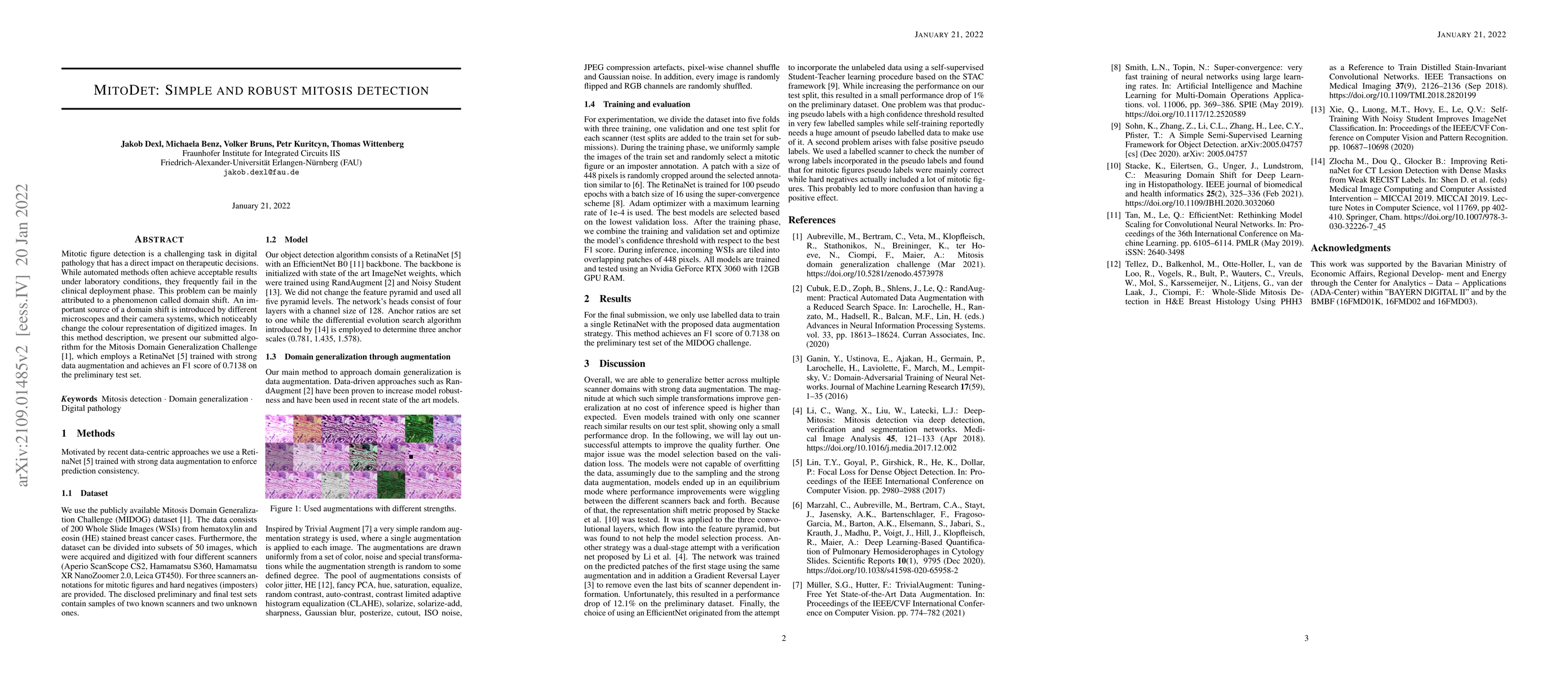
Discussion 0