Mixture theory modeling for characterizing solute transport in breast tumor tissues
Publication
Metrics
AI Quick Summary
This paper uses mixture theory to model solute transport in breast tumor tissues, comparing different vascular configurations. The study finds that dual capillaries and lymph vessels reduce extravascular solute concentration, with nonhomogeneous distributions for larger particles and local trapping for smaller ones. A universal non-dimensional time scale is identified for peak solute concentration, potentially aiding in therapeutic efficacy studies.
Paper Preview
Abstract
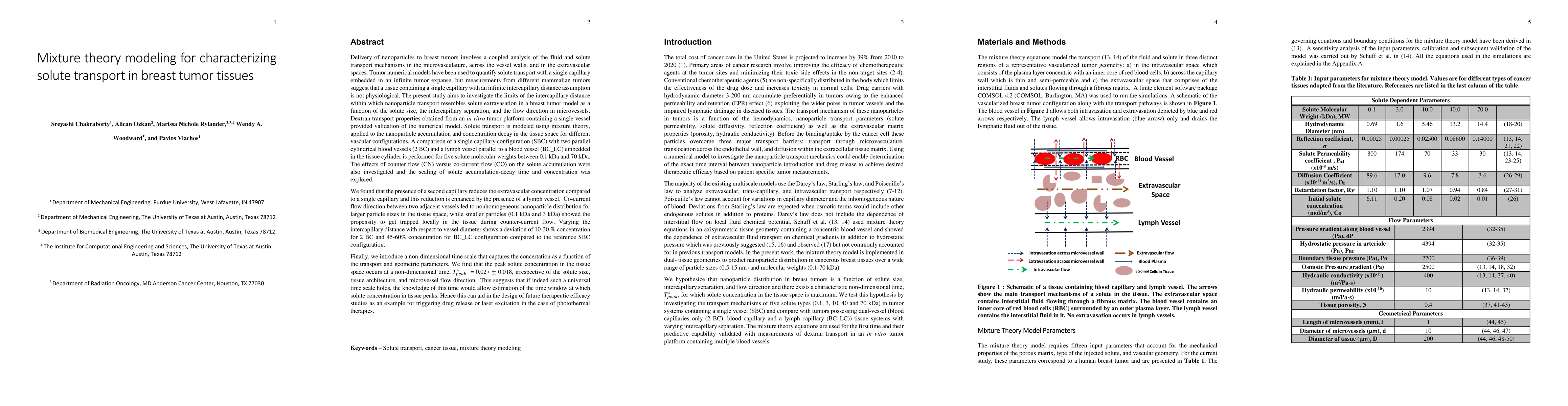
Solute transport is modeled using mixture theory, applied to the nanoparticle accumulation and concentration decay in the tissue space for different vascular configurations. A comparison of a single capillary configuration (SBC) with two parallel cylindrical blood vessels (2 BC) and a lymph vessel parallel to a blood vessel (BC_LC) embedded in the tissue cylinder is performed for five solute molecular weights between 0.1 kDa and 70 kDa. We found that the presence of a second capillary reduces the extravascular concentration compared to a single capillary and this reduction is enhanced by the presence of a lymph vessel. Co-current flow direction between two adjacent vessels led to nonhomogeneous nanoparticle distribution for larger particle sizes in the tissue space, while smaller particles (0.1 kDa and 3 kDa) showed the propensity to get trapped locally in the tissue during counter-current flow. Varying the intercapillary distance with respect to vessel diameter shows a deviation of 10-30 % concentration for 2 BC and 45-60% concentration for BC_LC configuration compared to the reference SBC configuration. Finally, we introduce a non-dimensional time scale that captures the concertation as a function of the transport and geometric parameters. We find that the peak solute concentration in the tissue space occurs at a non-dimensional time, T_peak^* = 0.027+/-0.018, irrespective of the solute size, tissue architecture, and microvessel flow direction. This suggests that if indeed such a universal time scale holds, the knowledge of this time would allow estimation of the time window at which solute concentration in tissue peaks. Hence this can aid in the design of future therapeutic efficacy studies as an example for triggering drug release or laser excitation in the case of photothermal therapies.
AI Key Findings
Get AI-generated insights about this paper's methodology, results, significance, and more — seven facets brought into focus.
Impact
Paper Details
PDF Preview
Key Terms
Citation Network
Current paper (gray), citations (green), references (blue)
Display is limited for performance on very large graphs.
Discussion 0