Moire structures in twisted bilayer graphene studied by transmission electron microscopy
Publication
Metrics
AI Quick Summary
This study explores moire structures in twisted bilayer graphene (TBG) using transmission electron microscopy (TEM), showing that low-energy electron diffraction (around 236 eV) reveals moire periodicity peaks not visible at higher TEM energies. Holography experiments reveal that variations in atomic stacking can influence transmitted wave intensity, emphasizing the importance of electron imaging techniques for graphene studies.
Paper Preview
Abstract
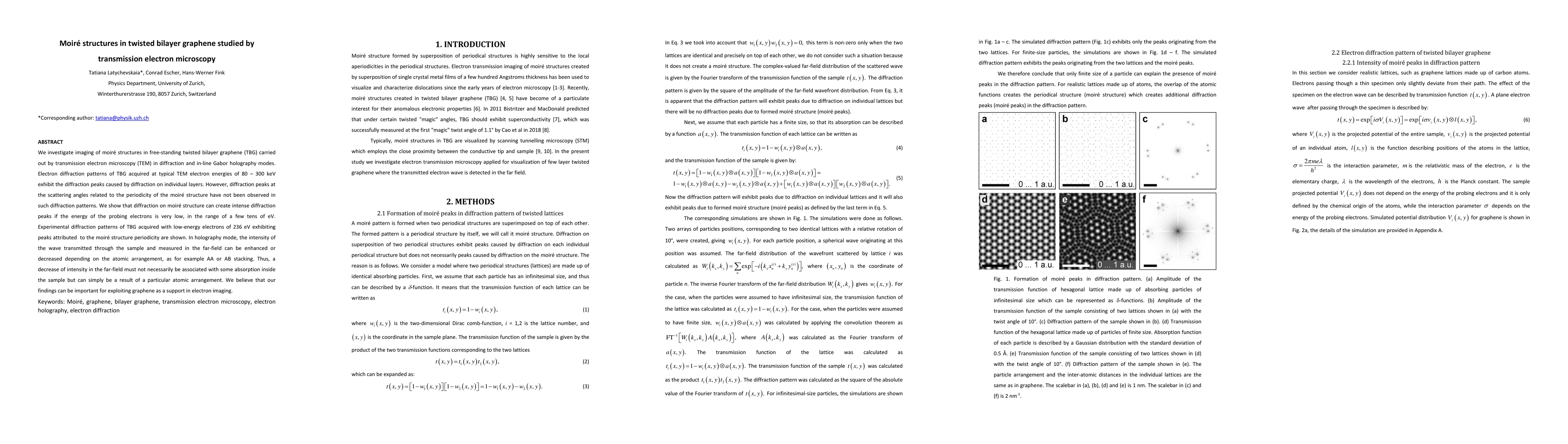
We investigate imaging of moire structures in free-standing twisted bilayer graphene (TBG) carried out by transmission electron microscopy (TEM) in diffraction and in-line Gabor holography modes. Electron diffraction patterns of TBG acquired at typical TEM electron energies of 80 - 300 keV exhibit the diffraction peaks caused by diffraction on individual layers. However, diffraction peaks at the scattering angles related to the periodicity of the moire structure have not been observed in such diffraction patterns. We show that diffraction on moire structure can create intense diffraction peaks if the energy of the probing electrons is very low, in the range of a few tens of eV. Experimental diffraction patterns of TBG acquired with low-energy electrons of 236 eV exhibiting peaks attributed to the moire structure periodicity are shown. In holography mode, the intensity of the wave transmitted through the sample and measured in the far-field can be enhanced or decreased depending on the atomic arrangement, as for example AA or AB stacking. Thus, a decrease of intensity in the far-field must not necessarily be associated with some absorption inside the sample but can simply be a result of a particular atomic arrangement. We believe that our findings can be important for exploiting graphene as a support in electron imaging.
AI Key Findings
Get AI-generated insights about this paper's methodology, results, significance, and more — seven facets brought into focus.
Impact
Paper Details
PDF Preview
Key Terms
Citation Network
Current paper (gray), citations (green), references (blue)
Display is limited for performance on very large graphs.
Discussion 0