ArxivLens

Quick Summary:

This paper employs advanced FRET biosensing and single-molecule optical microscopy to measure molecular crowding in single live yeast cells, revealing that crowding is influenced by extracellular ionic strength, local glucose conditions, and sensor copy number. The study demonstrates that cytosolic crowding is uniform and largely unaffected by cell membrane accessibility, providing insights into subcellular processes under stress.

arXiv Id: 2010.10862
Date Published: 2020-10-22
Date Updated: 2020-10-22

Categories:

Key Terms (TF-IDF):

cells (0.291)
cell (0.261)
molecule (0.248)
microscopy (0.237)
environment (0.187)
membrane (0.167)
copy (0.165)
quantification (0.160)
concentrations (0.155)
enabled (0.152)
brightness (0.148)
timescale (0.148)
track (0.144)
sensor (0.140)
single (0.140)
trajectories (0.138)
largely (0.138)
sensors (0.137)
stress (0.136)
life (0.135)
License: http://arxiv.org/licenses/nonexclusive-distrib/1.0/

Summary:

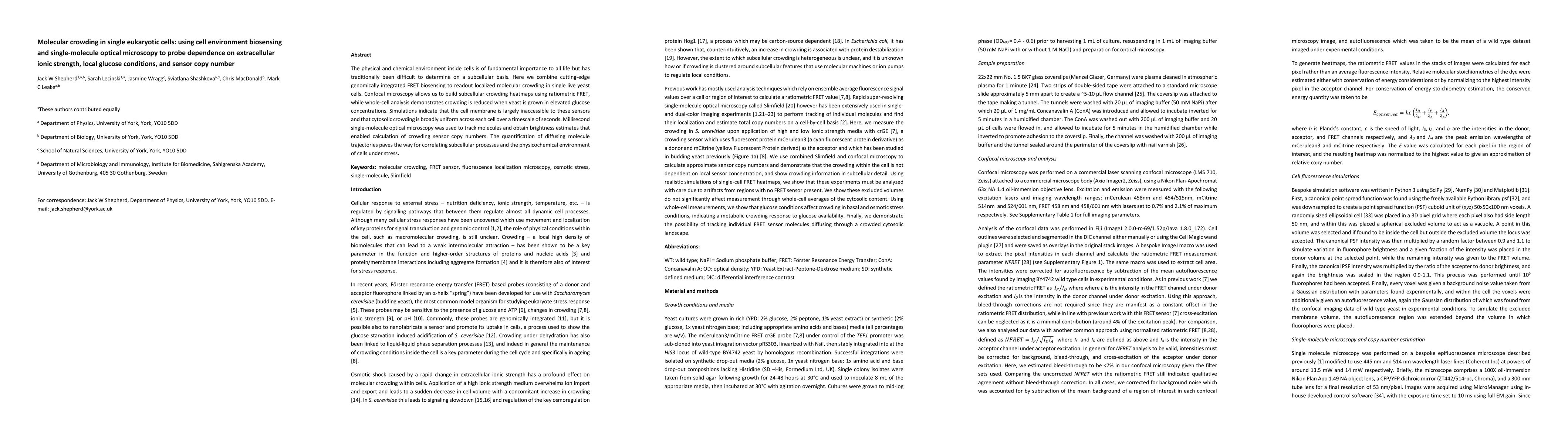
The physical and chemical environment inside cells is of fundamental importance to all life but has traditionally been difficult to determine on a subcellular basis. Here we combine cutting-edge genomically integrated FRET biosensing to readout localized molecular crowding in single live yeast cells. Confocal microscopy allows us to build subcellular crowding heatmaps using ratiometric FRET, while whole-cell analysis demonstrates crowding is reduced when yeast is grown in elevated glucose concentrations. Simulations indicate that the cell membrane is largely inaccessible to these sensors and that cytosolic crowding is broadly uniform across each cell over a timescale of seconds. Millisecond single-molecule optical microscopy was used to track molecules and obtain brightness estimates that enabled calculation of crowding sensor copy numbers. The quantification of diffusing molecule trajectories paves the way for correlating subcellular processes and the physicochemical environment of cells under stress.

Citation & Reference Network Visualization

This graph shows how this paper is connected to other research. The current paper is shown in dark gray, papers that cite it are in green, and papers it references are in blue. You can:

Current Paper
Papers citing this work (Citations)
Papers cited by this work (References)

• Hover over any node to see the paper title

• Click a node to view that paper's details

• Drag nodes to rearrange the graph

• Use the zoom controls (bottom right) to zoom in/out

Citations and References

Title Authors Year Actions
iPAR: a new reporter for eukaryotic cytoplasmic protein aggregation
Sarah Lecinski, Jamieson A L Howard, Chris MacDonald, M. Leake
2025
View Paper
Correlating viscosity and molecular crowding with fluorescent nanobeads and molecular probes: in vitro and in vivo
Sarah Lecinski, J. Shepherd, Kate R Bunting, Lara G. Dresser, Steven D. Quinn, C. MacDonald, M. Leake
2022
View Paper
Correlative approaches in single-molecule biophysics: a review of the progress in methods and applications.
M. Leake
2021
View Paper
PySTACHIO: Python Single-molecule TrAcking stoiCHiometry Intensity and simulatiOn, a flexible, extensible, beginner-friendly and optimized program for analysis of single-molecule microscopy data
J. Shepherd, E. Higgins, A. Wollman, M. Leake
2021
View Paper
Macromolecular Crowding: a Hidden Link Between Cell Volume and Everything Else.
M. Model, Jordan E. Hollembeak, M. Kurokawa
2021
View Paper
Investigating molecular crowding during cell division in budding yeast with FRET
Sarah Lecinski, J. Shepherd, Lewis Frame, Imogen Hayton, C. MacDonald, M. Leake
2021
View Paper
Localization microscopy: a review of the progress in methods and applications.
J. Shepherd, M. Leake
2020
View Paper

Comments (0)

This website uses cookies to ensure you get the best experience. Learn more