ArxivLens

Quick Summary:

This paper introduces MONAIfbs, a single-step UNet-based segmentation tool for fetal brain MRI in Spina Bifida, leveraging the MONAI framework. It outperforms a 2-step segmentation approach by achieving higher accuracy and reducing outliers, thus minimizing the need for manual corrections in Super Resolution Reconstruction algorithms.

arXiv Id: 2103.13314
Comments: Abstract accepted at IEEE International Symposium on Biomedical Imaging (ISBI) 2021
Date Published: 2021-03-25
Date Updated: 2021-03-25

Categories:

Key Terms (TF-IDF):

wang (0.291)
brain (0.275)
segmentation (0.256)
segmentation (0.255)
brain (0.250)
step (0.208)
step (0.199)
manual (0.178)
manual (0.174)
showed (0.159)
resolution (0.148)
showed (0.147)
resolution (0.145)
network (0.142)
network (0.138)
trained (0.128)
need (0.125)
need (0.124)
trained (0.124)
accurate (0.122)
License: http://arxiv.org/licenses/nonexclusive-distrib/1.0/

Summary:

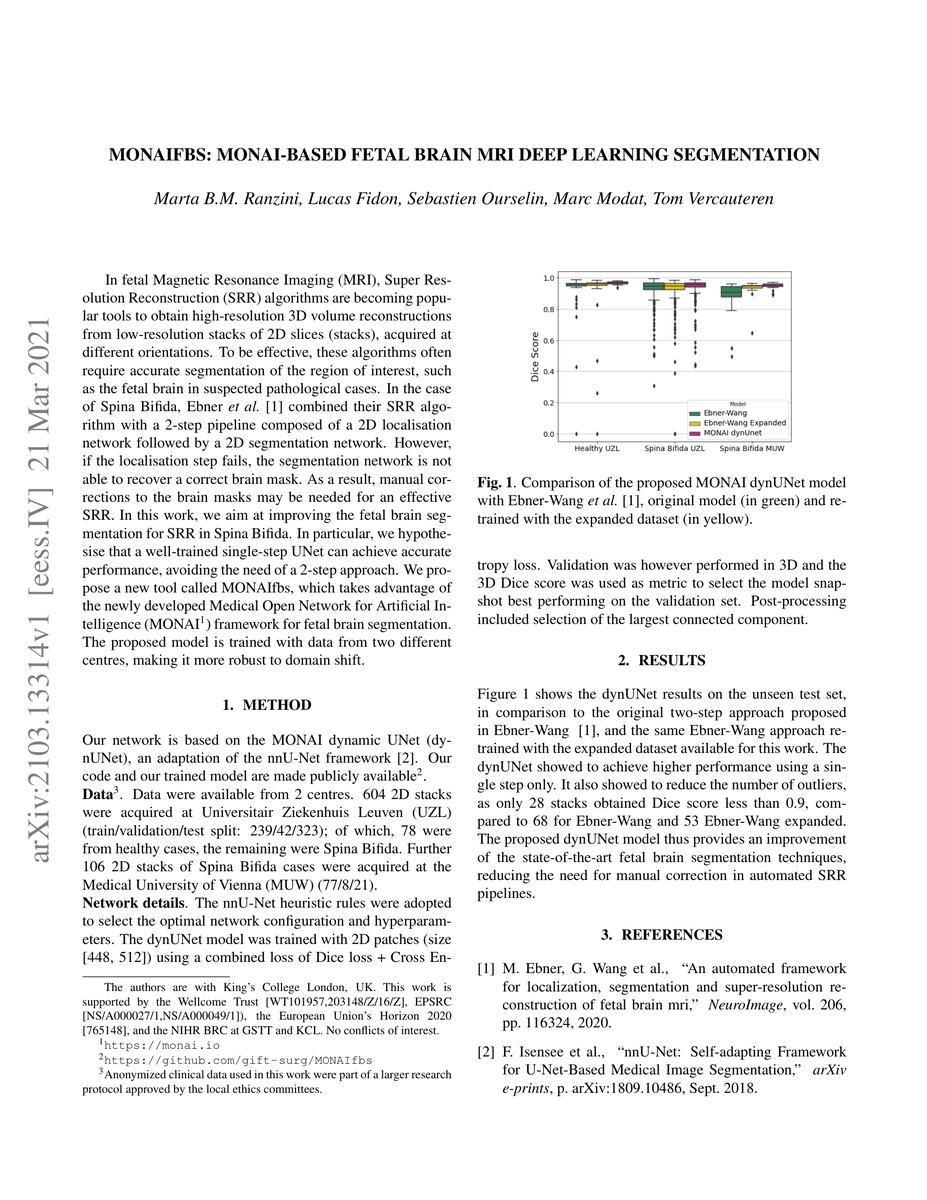
In fetal Magnetic Resonance Imaging, Super Resolution Reconstruction (SRR) algorithms are becoming popular tools to obtain high-resolution 3D volume reconstructions from low-resolution stacks of 2D slices, acquired at different orientations. To be effective, these algorithms often require accurate segmentation of the region of interest, such as the fetal brain in suspected pathological cases. In the case of Spina Bifida, Ebner, Wang et al. (NeuroImage, 2020) combined their SRR algorithm with a 2-step segmentation pipeline (2D localisation followed by a 2D segmentation network). However, if the localisation step fails, the second network is not able to recover a correct brain mask, thus requiring manual corrections for an effective SRR. In this work, we aim at improving the fetal brain segmentation for SRR in Spina Bifida. We hypothesise that a well-trained single-step UNet can achieve accurate performance, avoiding the need of a 2-step approach. We propose a new tool for fetal brain segmentation called MONAIfbs, which takes advantage of the Medical Open Network for Artificial Intelligence (MONAI) framework. Our network is based on the dynamic UNet (dynUNet), an adaptation of the nnU-Net framework. When compared to the original 2-step approach proposed in Ebner-Wang, and the same Ebner-Wang approach retrained with the expanded dataset available for this work, the dynUNet showed to achieve higher performance using a single step only. It also showed to reduce the number of outliers, as only 28 stacks obtained Dice score less than 0.9, compared to 68 for Ebner-Wang and 53 Ebner-Wang expanded. The proposed dynUNet model thus provides an improvement of the state-of-the-art fetal brain segmentation techniques, reducing the need for manual correction in automated SRR pipelines. Our code and our trained model are made publicly available at https://github.com/gift-surg/MONAIfbs.

Citation & Reference Network Visualization

This graph shows how this paper is connected to other research. The current paper is shown in dark gray, papers that cite it are in green, and papers it references are in blue. You can:

Current Paper
Papers citing this work (Citations)
Papers cited by this work (References)

• Hover over any node to see the paper title

• Click a node to view that paper's details

• Drag nodes to rearrange the graph

• Use the zoom controls (bottom right) to zoom in/out

Citations and References

Title Authors Year Actions
MorphiNet: A Graph Subdivision Network for Adaptive Bi-ventricle Surface Reconstruction
Yu Deng, Yiyang Xu, Linglong Qian, C. Mauger, Anastasia Nasopoulou, Steven Williams, Michelle C. Williams, Steven Niederer, David E. Newby, Andrew McCulloch, J. Omens, K. Pushparajah, Alistair A. Young
2024
View Paper
Automatic segmentation, classification, and prediction of pelvic bone tumors using deep learning techniques
Mona Shouman, K. Rahouma, Hesham F. A. Hamed
2024
View Paper
The Quest for the Application of Artificial Intelligence to Whole Slide Imaging: Unique Prospective from New Advanced Tools
Gavino Faa, Massimo Castagnola, Luca Didaci, Fernando Coghe, M. Scartozzi, Luca Saba, Matteo Fraschini
2024
View Paper
An Examination of Methodological Approaches for Segmentating Fetal Brain MRI Images – Analysis
Nilima Gaikwad, S. Vijayalakshmi
2024
View Paper
SurgNet: Self-Supervised Pretraining With Semantic Consistency for Vessel and Instrument Segmentation in Surgical Images
Jiachen Chen, Mengyang Li, Hu Han, Zhiming Zhao, Xilin Chen
2023
View Paper
FetMRQC: A robust quality control system for multi-centric fetal brain MRI
Thomas Sanchez, Oscar Esteban, Yvan Gomez, A. Pron, Mériam Koob, Vincent Dunet, Nadine Girard, A. Jakab, E. Eixarch, G. Auzias, M. Cuadra
2023
View Paper
Fetal-BET: Brain Extraction Tool for Fetal MRI
Razieh Faghihpirayesh, Davood Karimi, Deniz Erdoğmuş, A. Gholipour
2023
View Paper
Automatic Ventriculomegaly Detection in Fetal Brain MRI: A Step-by-Step Deep Learning Model for Novel 2D-3D Linear Measurements
Farzan Vahedifard, H. Ai, M. Supanich, Kranthi K. Marathu, Xuchu Liu, M. Koçak, Shehbaz M. Ansari, Melih Akyuz, Jubril O. Adepoju, Seth Adler, S. Byrd
2023
View Paper
FetMRQC: Automated Quality Control for fetal brain MRI
Thomas Sanchez, O. Esteban, Yvan Gomez, E. Eixarch, M. Cuadra
2023
View Paper
Continuous U-Net: Faster, Greater and Noiseless
Chunyang Cheng, Christina Runkel, Lihao Liu, R. Chan, C. Schonlieb, Angelica I. Avilés-Rivero
2023
View Paper
NeSVoR: Implicit Neural Representation for Slice-to-Volume Reconstruction in MRI
Junshen Xu, Daniel Moyer, B. Gagoski, J. E. Iglesias, P. Ellen Grant, P. Golland, E. Adalsteinsson
2023
View Paper
BEAN: Brain Extraction and Alignment Network for 3D Fetal Neurosonography
Felipe Moser, Ruobing Huang, INTERGROWTH-21 Consortium, B. Papież, Ana I. L. Namburete
2022
View Paper
A Dempster-Shafer Approach to Trustworthy AI With Application to Fetal Brain MRI Segmentation
L. Fidon, M. Aertsen, F. Kofler, A. Bink, A. David, T. Deprest, D. Emam, F. Guffens, A. Jakab, G. Kasprian, P. Kienast, A. Melbourne, Bjoern H Menze, N. Mufti, I. Pogledić, D. Prayer, Marlene Stuempflen, Esther Van Elslander, S. Ourselin, J. Deprest, Tom Kamiel Magda Vercauteren
2022
View Paper
Partial supervision for the FeTA challenge 2021
L. Fidon, M. Aertsen, Suprosanna Shit, P. Demaerel, S. Ourselin, J. Deprest, Tom Kamiel Magda Vercauteren
2021
View Paper
A spatio-temporal atlas of the developing fetal brain with spina bifida aperta
L. Fidon, Elizabeth Viola, N. Mufti, A. David, A. Melbourne, P. Demaerel, S. Ourselin, T. Vercauteren, J. Deprest, M. Aertsen
2021
View Paper
Distributionally Robust Segmentation of Abnormal Fetal Brain 3D MRI
L. Fidon, M. Aertsen, N. Mufti, T. Deprest, D. Emam, F. Guffens, E. Schwartz, M. Ebner, D. Prayer, G. Kasprian, A. David, A. Melbourne, S. Ourselin, J. Deprest, G. Langs, Tom Kamiel Magda Vercauteren
2021
View Paper
Label-set Loss Functions for Partial Supervision: Application to Fetal Brain 3D MRI Parcellation
L. Fidon, M. Aertsen, D. Emam, N. Mufti, F. Guffens, T. Deprest, P. Demaerel, A. David, A. Melbourne, S. Ourselin, Jam Deprest, Tom Kamiel Magda Vercauteren
2021
View Paper
TorchIO: A Python library for efficient loading, preprocessing, augmentation and patch-based sampling of medical images in deep learning
Fernando Pérez-García, R. Sparks, S. Ourselin
2020
View Paper
Distributionally Robust Deep Learning using Hardness Weighted Sampling
Lucas Fidon, S. Ourselin, T. Vercauteren
2020
View Paper
Journal Pre-proof Brain Extraction and Alignment Network for 3D Fetal Neurosonography
A. Namburete
2022
View Paper
Computer Methods and Programs in Biomedicine
Fernando Pérez-García, Rachel Sparks, Sébastien Ourselin
View Paper

Similar Papers

Comments (0)

This website uses cookies to ensure you get the best experience. Learn more