More Knowledge is Better: Cross-Modality Volume Completion and 3D+2D Segmentation for Intracardiac Echocardiography Contouring
Publication
Metrics
AI Quick Summary
This paper presents an automatic segmentation algorithm for intracardiac echocardiography (ICE) using a cross-modality framework that combines 3D CT data with ICE's 2D views, significantly improving the delineation of the left atrium and pulmonary veins. The method outperforms traditional 2D-only segmentation, especially for less-visible anatomical structures.
Paper Preview
Abstract
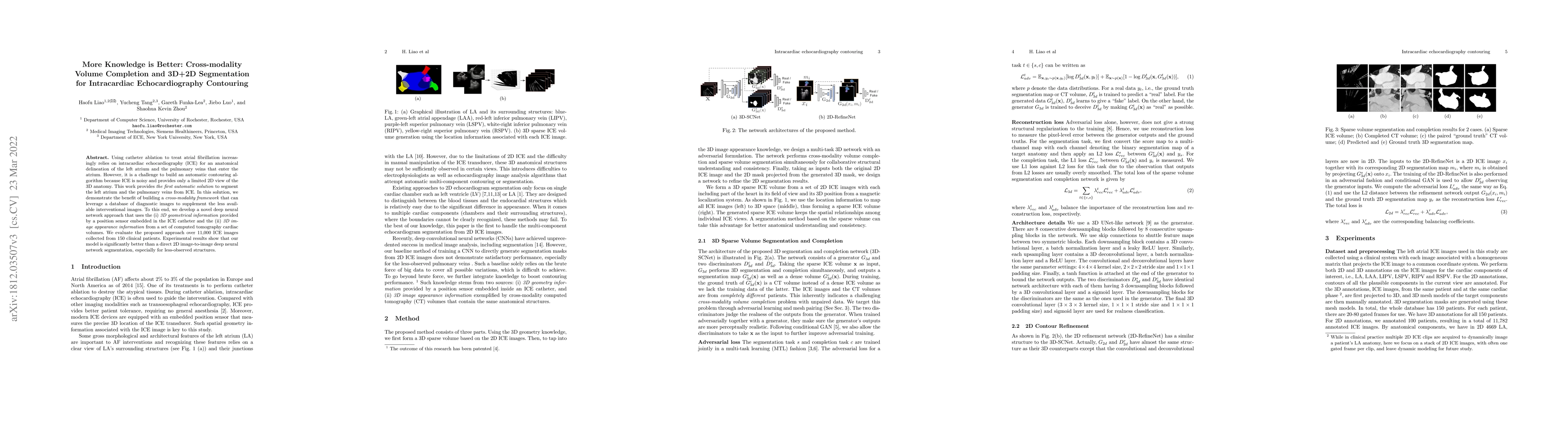
Using catheter ablation to treat atrial fibrillation increasingly relies on intracardiac echocardiography (ICE) for an anatomical delineation of the left atrium and the pulmonary veins that enter the atrium. However, it is a challenge to build an automatic contouring algorithm because ICE is noisy and provides only a limited 2D view of the 3D anatomy. This work provides the first automatic solution to segment the left atrium and the pulmonary veins from ICE. In this solution, we demonstrate the benefit of building a cross-modality framework that can leverage a database of diagnostic images to supplement the less available interventional images. To this end, we develop a novel deep neural network approach that uses the (i) 3D geometrical information provided by a position sensor embedded in the ICE catheter and the (ii) 3D image appearance information from a set of computed tomography cardiac volumes. We evaluate the proposed approach over 11,000 ICE images collected from 150 clinical patients. Experimental results show that our model is significantly better than a direct 2D image-to-image deep neural network segmentation, especially for less-observed structures.
AI Key Findings
Get AI-generated insights about this paper's methodology, results, significance, and more — seven facets brought into focus.
Impact
Paper Details
Authors
PDF Preview
Key Terms
Citation Network
Current paper (gray), citations (green), references (blue)
Display is limited for performance on very large graphs.
Discussion 0