MorphoITH: A Framework for Deconvolving Intra-Tumor Heterogeneity Using Tissue Morphology

Source: ArXiv

AI Quick Summary

MorphoITH is a novel framework that uses histopathology slides to deconvolve intra-tumor heterogeneity (ITH) through tissue morphology, offering a scalable alternative to resource-intensive sequencing methods. It captures clinically significant biological features and recognizes subclonal changes, bridging histopathology and genomics for precision oncology.

Share: Share on X LinkedIn

Summary

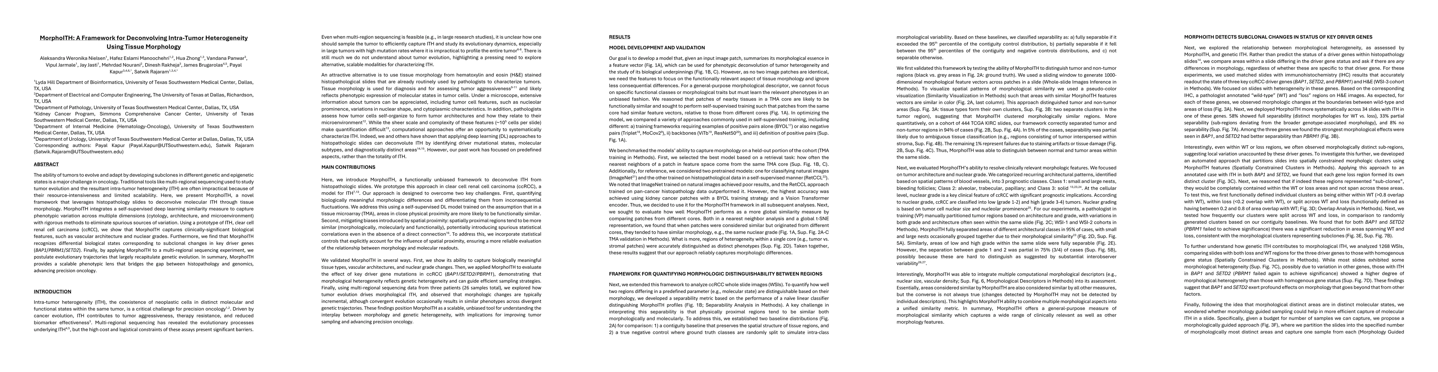
The ability of tumors to evolve and adapt by developing subclones in different genetic and epigenetic states is a major challenge in oncology. Traditional tools like multi-regional sequencing used to study tumor evolution and the resultant intra-tumor heterogeneity (ITH) are often impractical because of their resource-intensiveness and limited scalability. Here, we present MorphoITH, a novel framework that leverages histopathology slides to deconvolve molecular ITH through tissue morphology. MorphoITH integrates a self-supervised deep learning similarity measure to capture phenotypic variation across multiple dimensions (cytology, architecture, and microenvironment) with rigorous methods to eliminate spurious sources of variation. Using a prototype of ITH, clear cell renal cell carcinoma (ccRCC), we show that MorphoITH captures clinically-significant biological features, such as vascular architecture and nuclear grades. Furthermore, we find that MorphoITH recognizes differential biological states corresponding to subclonal changes in key driver genes (BAP1/PBRM1/SETD2). Finally, by applying MorphoITH to a multi-regional sequencing experiment, we postulate evolutionary trajectories that largely recapitulate genetic evolution. In summary, MorphoITH provides a scalable phenotypic lens that bridges the gap between histopathology and genomics, advancing precision oncology.

AI Key Findings

Generated Jun 12, 2025

Methodology

MorphoITH integrates a self-supervised deep learning similarity measure to capture phenotypic variation across cytology, architecture, and microenvironment, using histopathology slides to deconvolve molecular ITH through tissue morphology.

Key Results

  • MorphoITH captures clinically-significant biological features such as vascular architecture and nuclear grades in clear cell renal cell carcinoma (ccRCC).
  • MorphoITH recognizes differential biological states corresponding to subclonal changes in key driver genes (BAP1/PBRM1/SETD2).
  • MorphoITH postulates evolutionary trajectories that recapitulate genetic evolution when applied to a multi-regional sequencing experiment.

Significance

MorphoITH provides a scalable phenotypic lens that bridges the gap between histopathology and genomics, advancing precision oncology by offering a less resource-intensive approach to study tumor evolution and intra-tumor heterogeneity.

Technical Contribution

MorphoITH introduces a self-supervised deep learning framework that deconvolves molecular intra-tumor heterogeneity using tissue morphology from histopathology slides.

Novelty

MorphoITH distinguishes itself by leveraging tissue morphology and self-supervised deep learning to overcome the limitations of resource-intensive methods like multi-regional sequencing, offering a more scalable approach to study intra-tumor heterogeneity.

Limitations

  • The study was primarily validated on clear cell renal cell carcinoma (ccRCC), so further investigation is needed to confirm its applicability to other cancer types.
  • MorphoITH's performance might be influenced by the quality and resolution of histopathology slides.

Future Work

  • Explore the applicability of MorphoITH across diverse cancer types and tissue contexts.
  • Investigate the impact of histopathology slide quality and resolution on MorphoITH's performance.

Paper Details

PDF Preview

Download PDF

No citations found for this paper.

Comments (0)

This website uses cookies to ensure you get the best experience. Learn more