Morphology-Enhanced CAM-Guided SAM for weakly supervised Breast Lesion Segmentation
Publication
Metrics
AI Quick Summary
This paper proposes a novel weakly supervised framework using morphological enhancement, class activation maps, and the Segment Anything Model for segmenting breast lesions in ultrasound images, reducing the need for extensive manual labeling while achieving high segmentation accuracy comparable to supervised methods.
Paper Preview
Abstract
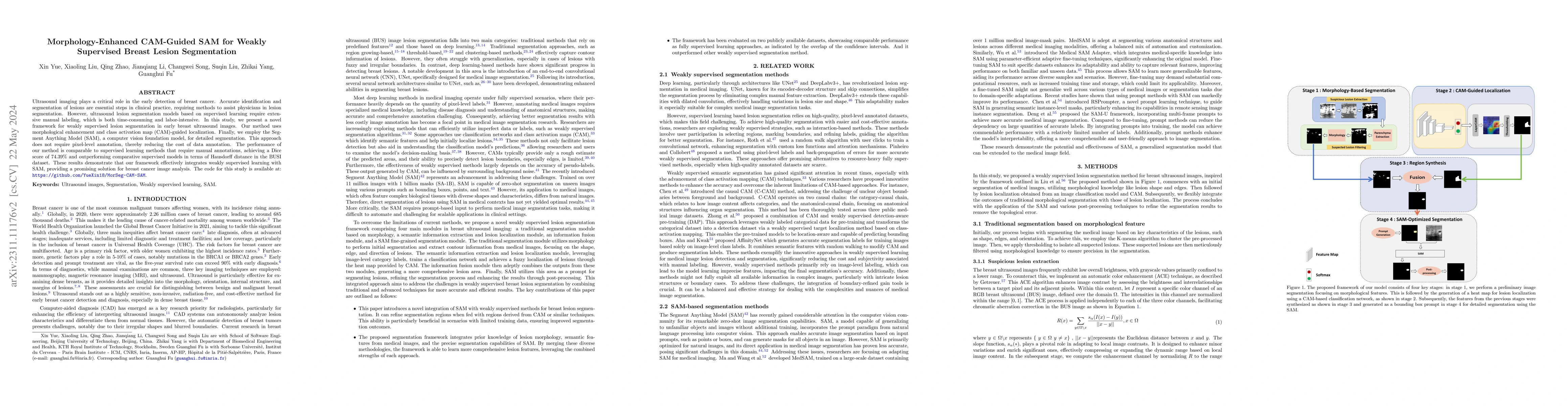
Ultrasound imaging plays a critical role in the early detection of breast cancer. Accurate identification and segmentation of lesions are essential steps in clinical practice, requiring methods to assist physicians in lesion segmentation. However, ultrasound lesion segmentation models based on supervised learning require extensive manual labeling, which is both time-consuming and labor-intensive. In this study, we present a novel framework for weakly supervised lesion segmentation in early breast ultrasound images. Our method uses morphological enhancement and class activation map (CAM)-guided localization. Finally, we employ the Segment Anything Model (SAM), a computer vision foundation model, for detailed segmentation. This approach does not require pixel-level annotation, thereby reducing the cost of data annotation. The performance of our method is comparable to supervised learning methods that require manual annotations, achieving a Dice score of 74.39% and outperforming comparative supervised models in terms of Hausdorff distance in the BUSI dataset. These results demonstrate that our framework effectively integrates weakly supervised learning with SAM, providing a promising solution for breast cancer image analysis. The code for this study is available at: https://github.com/YueXin18/MorSeg-CAM-SAM.
AI Key Findings
Get AI-generated insights about this paper's methodology, results, significance, and more — seven facets brought into focus.
Impact
Paper Details
Authors
PDF Preview
Key Terms
Citation Network
Current paper (gray), citations (green), references (blue)
Display is limited for performance on very large graphs.
Discussion 0