Publication
Metrics
Paper Preview
Abstract
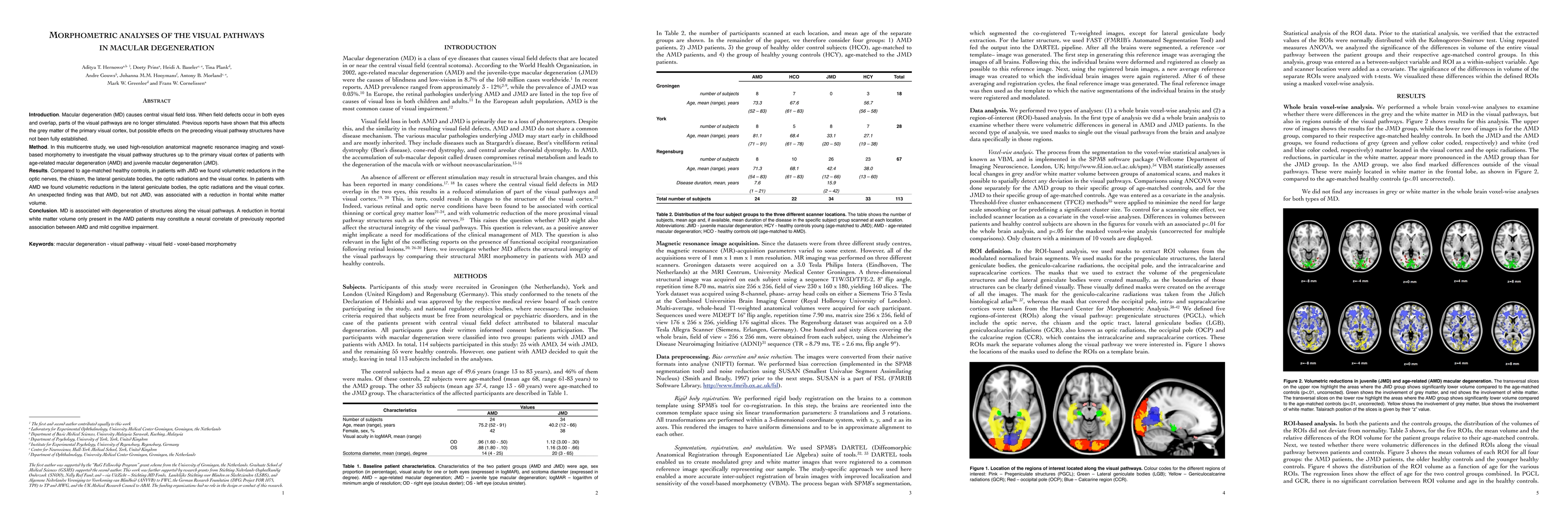
Introduction. Macular degeneration (MD) causes central visual field loss. When field defects occur in both eyes and overlap, parts of the visual pathways are no longer stimulated. Previous reports have shown that this affects the grey matter of the primary visual cortex, but possible effects on the preceding visual pathway structures have not been fully established. Method. In this multicentre study, we used high-resolution anatomical magnetic resonance imaging and voxel-based morphometry to investigate the visual pathway structures up to the primary visual cortex of patients with age-related macular degeneration (AMD) and juvenile macular degeneration (JMD). Results. Compared to age-matched healthy controls, in patients with JMD we found volumetric reductions in the optic nerves, the chiasm, the lateral geniculate bodies, the optic radiations and the visual cortex. In patients with AMD we found volumetric reductions in the lateral geniculate bodies, the optic radiations and the visual cortex. An unexpected finding was that AMD, but not JMD, was associated with a reduction in frontal white matter volume. Conclusion. MD is associated with degeneration of structures along the visual pathways. A reduction in frontal white matter volume only present in the AMD patients may constitute a neural correlate of previously reported association between AMD and mild cognitive impairment. Keywords: macular degeneration - visual pathway - visual field - voxel-based morphometry
AI Key Findings
Get AI-generated insights about this paper's methodology, results, significance, and more — seven facets brought into focus.
Impact
Paper Details
PDF Preview
Key Terms
Citation Network
Current paper (gray), citations (green), references (blue)
Display is limited for performance on very large graphs.
Discussion 0