MRI-based Head and Neck Tumor Segmentation Using nnU-Net with 15-fold Cross-Validation Ensemble
Publication
Metrics
AI Quick Summary
This study leverages MRI's superior soft tissue differentiation to improve head and neck tumor segmentation using nnU-Net with a 15-fold cross-validation ensemble, achieving an aggregated Dice Similarity Coefficient of 0.81 for pre-RT and 0.70 for mid-RT segmentation in a blind test set. The method's effectiveness is demonstrated through precise delineation of primary and metastatic lymph node tumors, potentially enhancing adaptive radiotherapy planning.
Paper Preview
Abstract
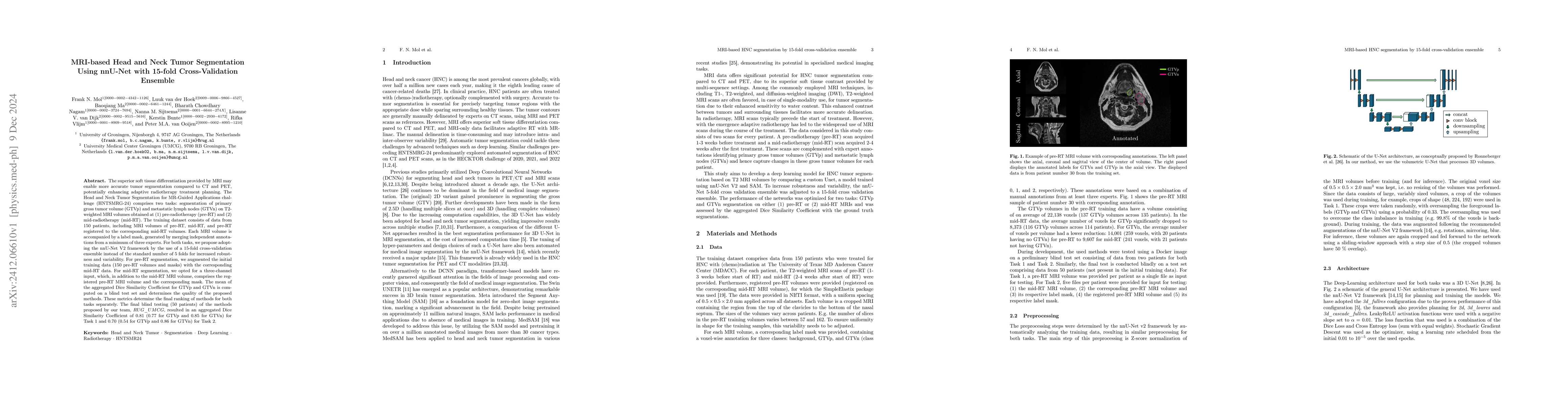
The superior soft tissue differentiation provided by MRI may enable more accurate tumor segmentation compared to CT and PET, potentially enhancing adaptive radiotherapy treatment planning. The Head and Neck Tumor Segmentation for MR-Guided Applications challenge (HNTSMRG-24) comprises two tasks: segmentation of primary gross tumor volume (GTVp) and metastatic lymph nodes (GTVn) on T2-weighted MRI volumes obtained at (1) pre-radiotherapy (pre-RT) and (2) mid-radiotherapy (mid-RT). The training dataset consists of data from 150 patients, including MRI volumes of pre-RT, mid-RT, and pre-RT registered to the corresponding mid-RT volumes. Each MRI volume is accompanied by a label mask, generated by merging independent annotations from a minimum of three experts. For both tasks, we propose adopting the nnU-Net V2 framework by the use of a 15-fold cross-validation ensemble instead of the standard number of 5 folds for increased robustness and variability. For pre-RT segmentation, we augmented the initial training data (150 pre-RT volumes and masks) with the corresponding mid-RT data. For mid-RT segmentation, we opted for a three-channel input, which, in addition to the mid-RT MRI volume, comprises the registered pre-RT MRI volume and the corresponding mask. The mean of the aggregated Dice Similarity Coefficient for GTVp and GTVn is computed on a blind test set and determines the quality of the proposed methods. These metrics determine the final ranking of methods for both tasks separately. The final blind testing (50 patients) of the methods proposed by our team, RUG_UMCG, resulted in an aggregated Dice Similarity Coefficient of 0.81 (0.77 for GTVp and 0.85 for GTVn) for Task 1 and 0.70 (0.54 for GTVp and 0.86 for GTVn) for Task 2.
AI Key Findings
Get AI-generated insights about this paper's methodology, results, significance, and more — seven facets brought into focus.
Impact
Authors
PDF Preview
Citation Network
Current paper (gray), citations (green), references (blue)
Display is limited for performance on very large graphs.
Discussion 0