MRI Brain Tumor Segmentation using Random Forests and Fully Convolutional Networks
Publication
Metrics
AI Quick Summary
This paper presents an automated brain tumor segmentation method using a combination of fully convolutional networks and random forests, achieving promising results with Dice overlap measures of 0.86, 0.78, and 0.66 for whole, core, and enhancing tumors respectively, evaluated on the BRATS 2017 dataset.
Paper Preview
Abstract
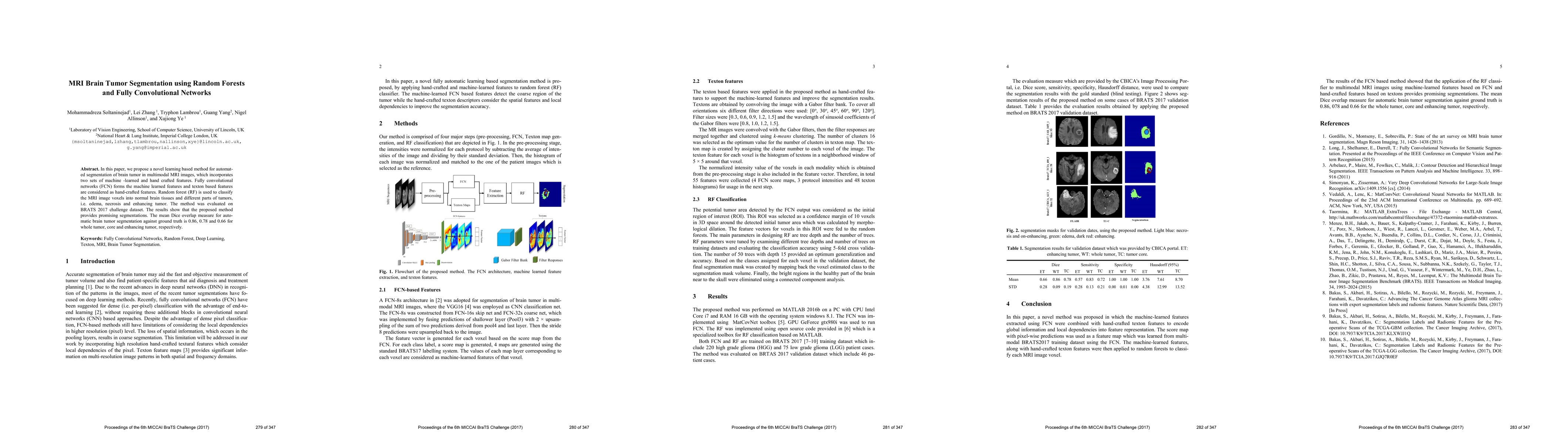
In this paper, we propose a novel learning based method for automated segmentation of brain tumor in multimodal MRI images, which incorporates two sets of machine -learned and hand crafted features. Fully convolutional networks (FCN) forms the machine learned features and texton based features are considered as hand-crafted features. Random forest (RF) is used to classify the MRI image voxels into normal brain tissues and different parts of tumors, i.e. edema, necrosis and enhancing tumor. The method was evaluated on BRATS 2017 challenge dataset. The results show that the proposed method provides promising segmentations. The mean Dice overlap measure for automatic brain tumor segmentation against ground truth is 0.86, 0.78 and 0.66 for whole tumor, core and enhancing tumor, respectively.
AI Key Findings
Get AI-generated insights about this paper's methodology, results, significance, and more — seven facets brought into focus.
Impact
Paper Details
Authors
PDF Preview
Key Terms
Citation Network
Current paper (gray), citations (green), references (blue)
Display is limited for performance on very large graphs.
Discussion 0