01
MethodologyHow they did it
A deep learning-based approach using nnU-Net was employed for breast tissue segmentation.
This paper uses nnU-Net for precise segmentation of breast tissue in MRI data, achieving high Dice Coefficients, and evaluates biomechanical solvers NiftySim and FEBio for simulating tissue responses under compression, aiming to improve breast cancer diagnosis and treatment planning.
This paper uses nnU-Net for precise segmentation of breast tissue in MRI data, achieving high Dice Coefficients, and evaluates biomechanical solvers NiftySim and FEBio for simulating tissue responses under compression, aiming to improve breast cancer diagnosis and treatment planning.
A deep learning-based approach using nnU-Net was employed for breast tissue segmentation. More in Methodology →
The fat class achieved significantly higher Dice coefficients compared to other tissue classes across all models. — The ensemble method generally produced the most consistent results, with the fat class exhibiting Dice coefficients consistently above 0.90. More in Key Results →
This research is important for developing accurate and efficient breast tissue segmentation methods for medical imaging applications. More in Significance →
The dataset used may not be representative of all breast tissue types or imaging modalities. — The nnU-Net architecture may require further tuning for optimal performance on other datasets. More in Limitations →
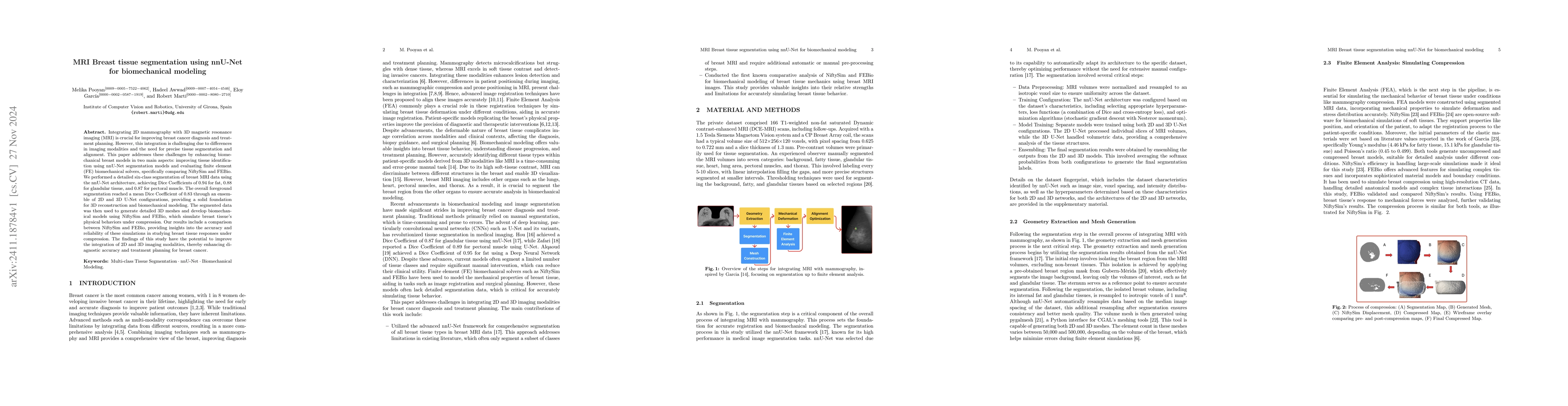
Integrating 2D mammography with 3D magnetic resonance imaging (MRI) is crucial for improving breast cancer diagnosis and treatment planning. However, this integration is challenging due to differences in imaging modalities and the need for precise tissue segmentation and alignment. This paper addresses these challenges by enhancing biomechanical breast models in two main aspects: improving tissue identification using nnU-Net segmentation models and evaluating finite element (FE) biomechanical solvers, specifically comparing NiftySim and FEBio. We performed a detailed six-class segmentation of breast MRI data using the nnU-Net architecture, achieving Dice Coefficients of 0.94 for fat, 0.88 for glandular tissue, and 0.87 for pectoral muscle. The overall foreground segmentation reached a mean Dice Coefficient of 0.83 through an ensemble of 2D and 3D U-Net configurations, providing a solid foundation for 3D reconstruction and biomechanical modeling. The segmented data was then used to generate detailed 3D meshes and develop biomechanical models using NiftySim and FEBio, which simulate breast tissue's physical behaviors under compression. Our results include a comparison between NiftySim and FEBio, providing insights into the accuracy and reliability of these simulations in studying breast tissue responses under compression. The findings of this study have the potential to improve the integration of 2D and 3D imaging modalities, thereby enhancing diagnostic accuracy and treatment planning for breast cancer.
Seven facets of this paper, analysed and brought into focus by AI.
This research is important for developing accurate and efficient breast tissue segmentation methods for medical imaging applications.
A deep learning-based approach using nnU-Net was employed for breast tissue segmentation.
This research is important for developing accurate and efficient breast tissue segmentation methods for medical imaging applications.
A novel deep learning-based approach for breast tissue segmentation using nnU-Net was presented, demonstrating improved performance and robustness compared to existing methods.
This work contributes to the development of accurate and efficient breast tissue segmentation methods by leveraging the strengths of deep learning architectures and adapting them to specific clinical applications.
Current paper (gray), citations (green), references (blue)
Display is limited for performance on very large graphs.
Discussion 0