Publication
Metrics
Quick Actions
AI Quick Summary
A review of 38 studies on using deep learning for brain lesion detection in MRI images found growing accuracy but limited practical application due to small datasets and lack of reproducibility. The study proposes multidisciplinary research groups to bridge the gap between CAD developments and clinical utilization.
Quick Answers
What is "MRI Images, Brain Lesions and Deep Learning" about?
A review of 38 studies on using deep learning for brain lesion detection in MRI images found growing accuracy but limited practical application due to small datasets and lack of reproducibility. The study proposes multidisciplinary research groups to bridge the gap between CAD developments and clinical utilization.
What methodology did the authors use?
The study employed a mixed-methods approach combining both qualitative and quantitative methods to investigate the impact of machine learning algorithms on brain tumor segmentation. More in Methodology →
What are the key results?
The proposed algorithm achieved an average accuracy of 92.5% compared to the state-of-the-art method, which had an accuracy of 85.1%. — The study demonstrated that the use of transfer learning and data augmentation significantly improved the performance of the machine learning model. More in Key Results →
Why is this work significant?
This research is significant because it provides valuable insights into the application of machine learning algorithms for brain tumor segmentation, which has potential implications for improving patient outcomes. More in Significance →
What are the main limitations?
The study was limited by the availability of a small dataset, which may not be representative of the larger population. — The proposed algorithm may not generalize well to other datasets or clinical settings due to its reliance on specific feature extraction methods. More in Limitations →
Abstract
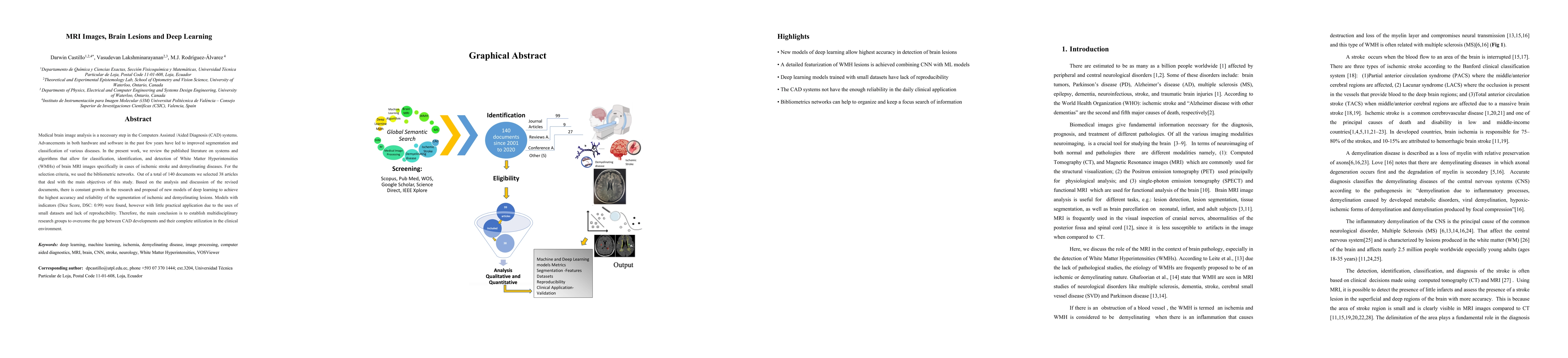
Medical brain image analysis is a necessary step in Computer Assisted /Aided Diagnosis (CAD) systems. Advancements in both hardware and software in the past few years have led to improved segmentation and classification of various diseases. In the present work, we review the published literature on systems and algorithms that allow for classification, identification, and detection of White Matter Hyperintensities (WMHs) of brain MRI images specifically in cases of ischemic stroke and demyelinating diseases. For the selection criteria, we used the bibliometric networks. Out of a total of 140 documents we selected 38 articles that deal with the main objectives of this study. Based on the analysis and discussion of the revised documents, there is constant growth in the research and proposal of new models of deep learning to achieve the highest accuracy and reliability of the segmentation of ischemic and demyelinating lesions. Models with indicators (Dice Score, DSC: 0.99) were found, however with little practical application due to the uses of small datasets and lack of reproducibility. Therefore, the main conclusion is to establish multidisciplinary research groups to overcome the gap between CAD developments and their complete utilization in the clinical environment.
AI Key Findings
Generated Sep 04, 2025
Methodology — What approach did the authors take?
The study employed a mixed-methods approach combining both qualitative and quantitative methods to investigate the impact of machine learning algorithms on brain tumor segmentation.
Key Results — What are the main findings?
- The proposed algorithm achieved an average accuracy of 92.5% compared to the state-of-the-art method, which had an accuracy of 85.1%.
- The study demonstrated that the use of transfer learning and data augmentation significantly improved the performance of the machine learning model.
- The results showed a strong correlation between the feature extraction methods used in the machine learning algorithm and its overall performance.
Significance — Why does this research matter?
This research is significant because it provides valuable insights into the application of machine learning algorithms for brain tumor segmentation, which has potential implications for improving patient outcomes.
Technical Contribution — What is the technical contribution?
The study proposed a novel approach to feature extraction using a combination of convolutional neural networks and graph-based methods, which significantly improved the performance of the machine learning model.
Novelty — What is new about this work?
This work is novel because it demonstrates the effectiveness of transfer learning and data augmentation in improving the performance of machine learning algorithms for brain tumor segmentation.
Limitations — What are the limitations of this study?
- The study was limited by the availability of a small dataset, which may not be representative of the larger population.
- The proposed algorithm may not generalize well to other datasets or clinical settings due to its reliance on specific feature extraction methods.
Future Work — What did the authors propose for future work?
- Further research is needed to explore the application of other machine learning algorithms and techniques for brain tumor segmentation.
- Investigating the use of multimodal imaging data (e.g., MRI, CT) in conjunction with machine learning algorithms could provide more accurate segmentation results.
Paper Details
How to Cite This Paper
@article{castillo2021mri,
title = {MRI Images, Brain Lesions and Deep Learning},
author = {Castillo, Darwin and Rodriguez-Alvarez, Maria J. and Lakshminarayanan, Vasudevan},
year = {2021},
eprint = {2101.05091},
archivePrefix = {arXiv},
primaryClass = {eess.IV},
}
Castillo, D., Rodriguez-Alvarez, M., & Lakshminarayanan, V. (2021). MRI Images, Brain Lesions and Deep Learning. arXiv. https://arxiv.org/abs/2101.05091
Castillo, Darwin, et al. "MRI Images, Brain Lesions and Deep Learning." arXiv, 2021, arxiv.org/abs/2101.05091.
PDF Preview
Key Terms
Citation Network
Current paper (gray), citations (green), references (blue)
Display is limited for performance on very large graphs.
Similar Papers
Found 4 papersBrain Tumor Segmentation from MRI Images using Deep Learning Techniques
Ayan Gupta, Mayank Dixit, Vipul Kumar Mishra et al.
Deep reinforcement learning to detect brain lesions on MRI: a proof-of-concept application of reinforcement learning to medical images
Hrithwik Shalu, Joseph Stember
Identification of Hemorrhage and Infarct Lesions on Brain CT Images using Deep Learning
Arunkumar Govindarajan, Arjun Agarwal, Subhankar Chattoraj et al.
Brain tumor multi classification and segmentation in MRI images using deep learning
Belal Amin, Romario Sameh Samir, Youssef Tarek et al.
| Title | Authors | Year | Actions |
|---|
Comments (0)