The automatic diagnosis of Parkinson's disease is in high clinical demand due
to its prevalence and the importance of targeted treatment. Current clinical
practice often relies on diagnostic biomarkers in QSM and NM-MRI images.
However, the lack of large, high-quality datasets makes training diagnostic
models from scratch prone to overfitting. Adapting pre-trained 3D medical
models is also challenging, as the diversity of medical imaging leads to
mismatches in voxel spacing and modality between pre-training and fine-tuning
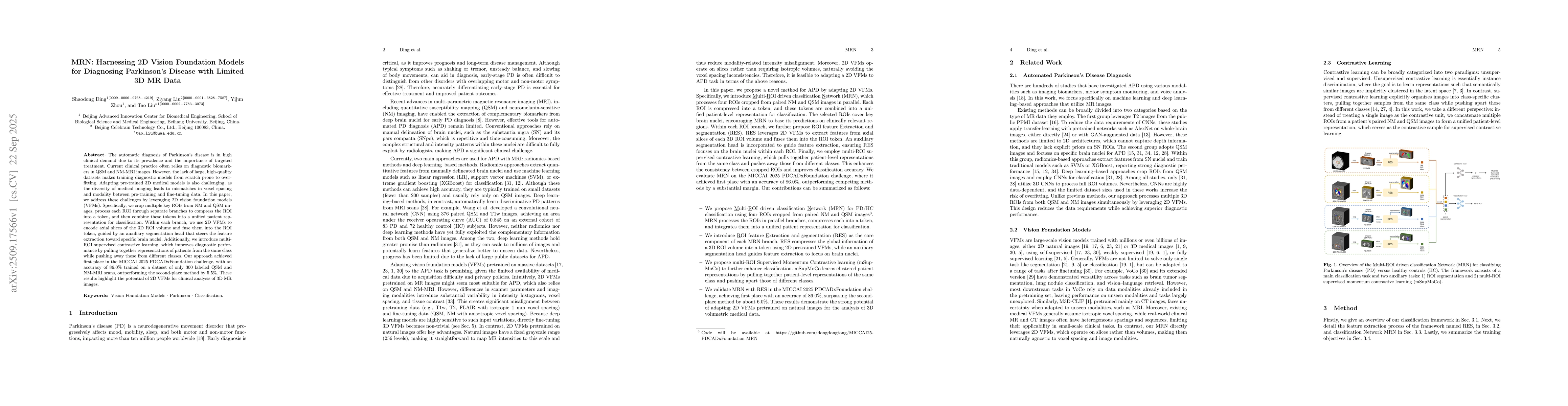
data. In this paper, we address these challenges by leveraging 2D vision
foundation models (VFMs). Specifically, we crop multiple key ROIs from NM and
QSM images, process each ROI through separate branches to compress the ROI into
a token, and then combine these tokens into a unified patient representation
for classification. Within each branch, we use 2D VFMs to encode axial slices
of the 3D ROI volume and fuse them into the ROI token, guided by an auxiliary
segmentation head that steers the feature extraction toward specific brain
nuclei. Additionally, we introduce multi-ROI supervised contrastive learning,
which improves diagnostic performance by pulling together representations of
patients from the same class while pushing away those from different classes.
Our approach achieved first place in the MICCAI 2025 PDCADxFoundation
challenge, with an accuracy of 86.0% trained on a dataset of only 300 labeled
QSM and NM-MRI scans, outperforming the second-place method by 5.5%.These
results highlight the potential of 2D VFMs for clinical analysis of 3D MR
images.
Discussion 0