Authors
Publication
Metrics
Quick Actions
Quick Answers
What methodology did the authors use?
The research introduces MSDM, a multimodal conditioned diffusion model that generates synthetic pathology images by integrating horizontal/vertical maps, RGB color characteristics, and BERT-encoded metadata through multi-head cross-attention mechanisms. More in Methodology →
What are the key results?
MSDM significantly reduces Wasserstein distances between real and synthetic images, demonstrating high fidelity in capturing biological conditions. — Synthetic data generated by MSDM improves segmentation accuracy for columnar cells compared to both real data and SDM-generated data. More in Key Results →
Why is this work significant?
This research addresses data scarcity in computational pathology by creating synthetic datasets that enhance model robustness and generalizability, enabling better segmentation of rare cell morphologies. More in Significance →
What are the main limitations?
The approach requires an initial set of annotated masks for challenging morphologies, which may not fully eliminate the need for manual annotation. — The effectiveness of synthetic data may vary depending on the specific morphological features and assay types. More in Limitations →
Abstract
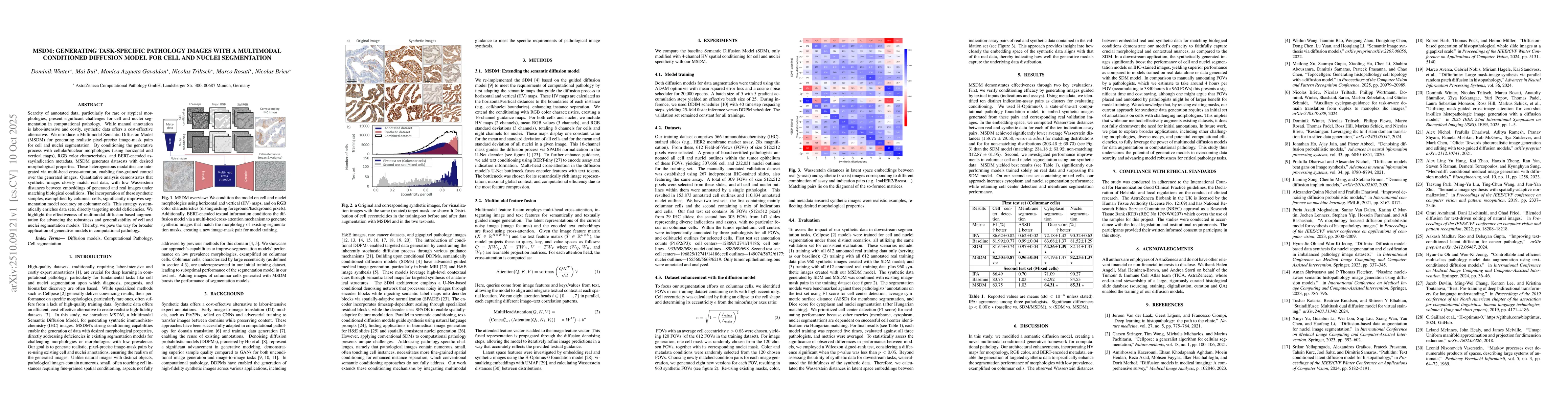
Scarcity of annotated data, particularly for rare or atypical morphologies, present significant challenges for cell and nuclei segmentation in computational pathology. While manual annotation is labor-intensive and costly, synthetic data offers a cost-effective alternative. We introduce a Multimodal Semantic Diffusion Model (MSDM) for generating realistic pixel-precise image-mask pairs for cell and nuclei segmentation. By conditioning the generative process with cellular/nuclear morphologies (using horizontal and vertical maps), RGB color characteristics, and BERT-encoded assay/indication metadata, MSDM generates datasests with desired morphological properties. These heterogeneous modalities are integrated via multi-head cross-attention, enabling fine-grained control over the generated images. Quantitative analysis demonstrates that synthetic images closely match real data, with low Wasserstein distances between embeddings of generated and real images under matching biological conditions. The incorporation of these synthetic samples, exemplified by columnar cells, significantly improves segmentation model accuracy on columnar cells. This strategy systematically enriches data sets, directly targeting model deficiencies. We highlight the effectiveness of multimodal diffusion-based augmentation for advancing the robustness and generalizability of cell and nuclei segmentation models. Thereby, we pave the way for broader application of generative models in computational pathology.
AI Key Findings
Generated Oct 16, 2025
Methodology — What approach did the authors take?
The research introduces MSDM, a multimodal conditioned diffusion model that generates synthetic pathology images by integrating horizontal/vertical maps, RGB color characteristics, and BERT-encoded metadata through multi-head cross-attention mechanisms.
Key Results — What are the main findings?
- MSDM significantly reduces Wasserstein distances between real and synthetic images, demonstrating high fidelity in capturing biological conditions.
- Synthetic data generated by MSDM improves segmentation accuracy for columnar cells compared to both real data and SDM-generated data.
- The model achieves better performance in cytoplasm and nucleus segmentation while maintaining cell center detection and membrane segmentation on mixed cell datasets.
Significance — Why does this research matter?
This research addresses data scarcity in computational pathology by creating synthetic datasets that enhance model robustness and generalizability, enabling better segmentation of rare cell morphologies.
Technical Contribution — What is the technical contribution?
The integration of multimodal conditioning (HV maps, RGB color, BERT metadata) with multi-head cross-attention in a diffusion model framework for generating task-specific pathology images.
Novelty — What is new about this work?
MSDM uniquely combines spatial, color, and textual modalities through cross-attention mechanisms, enabling fine-grained control over synthetic image generation for cell and nuclei segmentation in computational pathology.
Limitations — What are the limitations of this study?
- The approach requires an initial set of annotated masks for challenging morphologies, which may not fully eliminate the need for manual annotation.
- The effectiveness of synthetic data may vary depending on the specific morphological features and assay types.
Future Work — What did the authors propose for future work?
- Exploring broader applications with diverse morphologies, assays, and computational efficiencies.
- Investigating automated annotation techniques to reduce reliance on initial manual annotations.
- Enhancing multimodal conditioning to incorporate additional biological features for more precise synthesis.
How to Cite This Paper
@article{winter2025msdm,
title = {MSDM: Generating Task-Specific Pathology Images with a Multimodal
Conditioned Diffusion Model for Cell and Nuclei Segmentation},
author = {Winter, Dominik and Bui, Mai and Rosati, Marco and others},
year = {2025},
eprint = {2510.09121},
archivePrefix = {arXiv},
primaryClass = {cs.CV},
}
Winter, D., Bui, M., Rosati, M., Gavaldon, M., Brieu, N., & Triltsch, N. (2025). MSDM: Generating Task-Specific Pathology Images with a Multimodal
Conditioned Diffusion Model for Cell and Nuclei Segmentation. arXiv. https://arxiv.org/abs/2510.09121
Winter, Dominik, et al. "MSDM: Generating Task-Specific Pathology Images with a Multimodal
Conditioned Diffusion Model for Cell and Nuclei Segmentation." arXiv, 2025, arxiv.org/abs/2510.09121.
PDF Preview
Similar Papers
Found 4 papersMask-conditioned latent diffusion for generating gastrointestinal polyp images
Leila Mozaffari, Zahra Sepasdar, Sravanthi Parasa et al.
TopoCellGen: Generating Histopathology Cell Topology with a Diffusion Model
Meilong Xu, Saumya Gupta, Xiaoling Hu et al.
DiffMix: Diffusion Model-based Data Synthesis for Nuclei Segmentation and Classification in Imbalanced Pathology Image Datasets
Hyun-Jic Oh, Won-Ki Jeong
An Auxiliary Task for Learning Nuclei Segmentation in 3D Microscopy Images
Peter Hirsch, Dagmar Kainmueller
Comments (0)