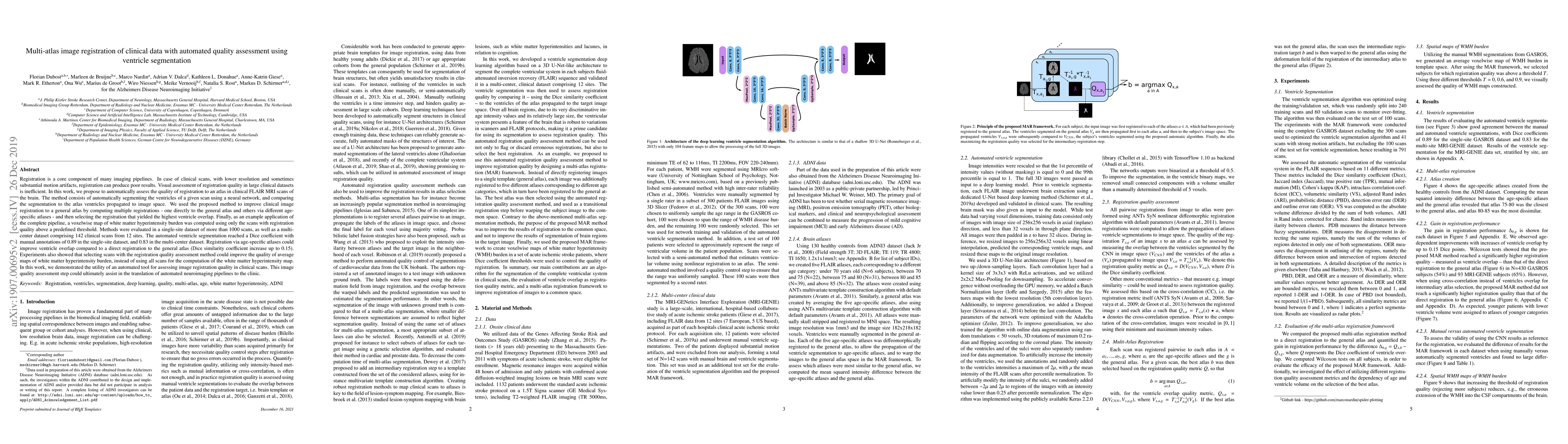
Quick Summary:

A new method automates quality assessment of brain MRI scans by segmenting ventricles and comparing to an atlas, improving image registration accuracy in clinical scans.

Summary

Registration is a core component of many imaging pipelines. In case of clinical scans, with lower resolution and sometimes substantial motion artifacts, registration can produce poor results. Visual assessment of registration quality in large clinical datasets is inefficient. In this work, we propose to automatically assess the quality of registration to an atlas in clinical FLAIR MRI scans of the brain. The method consists of automatically segmenting the ventricles of a given scan using a neural network, and comparing the segmentation to the atlas' ventricles propagated to image space. We used the proposed method to improve clinical image registration to a general atlas by computing multiple registrations and then selecting the registration that yielded the highest ventricle overlap. Methods were evaluated in a single-site dataset of more than 1000 scans, as well as a multi-center dataset comprising 142 clinical scans from 12 sites. The automated ventricle segmentation reached a Dice coefficient with manual annotations of 0.89 in the single-site dataset, and 0.83 in the multi-center dataset. Registration via age-specific atlases could improve ventricle overlap compared to a direct registration to the general atlas (Dice similarity coefficient increase up to 0.15). Experiments also showed that selecting scans with the registration quality assessment method could improve the quality of average maps of white matter hyperintensity burden, instead of using all scans for the computation of the white matter hyperintensity map. In this work, we demonstrated the utility of an automated tool for assessing image registration quality in clinical scans. This image quality assessment step could ultimately assist in the translation of automated neuroimaging pipelines to the clinic.

AI Key Findings

Get AI-generated insights about this paper's methodology, results, and significance.

Paper Details

PDF Preview

Download PDF

Key Terms

clinical (0.284)
atlas (0.268)
quality (0.204)
assessment (0.204)
automated (0.202)
overlap (0.174)
dataset (0.171)
selecting (0.169)
site (0.168)
image (0.167)
white (0.162)
segmentation (0.155)
automatically (0.154)
improve (0.147)
coefficient (0.143)
center (0.139)
matter (0.118)
scan (0.112)
reached (0.112)
annotations (0.111)
Title Authors Year Actions
Cardiac cavity segmentation review in the past decade: Methods and future perspectives
Feiyan Li, Weisheng Li, Yucheng Shu, Yidong Peng, Bin Xiao
2025
View Paper
Automated ventricular segmentation and shunt failure detection using convolutional neural networks
Kevin T. Huang, Jack McNulty, Helweh Hussein, Neil Klinger, M. Chua, Patrick R. Ng, J. Chalif, Neel H. Mehta, Omar Arnaout
2024
View Paper
Assessing CT-based Volumetric Analysis via Transfer Learning with MRI and Manual Labels for Idiopathic Normal Pressure Hydrocephalus
M. Srikrishna, Woosung Seo, A. Zettergren, S. Kern, Daniel Cantré, F. Gessler, H. Sotoudeh, Jakob Seidlitz, J. Bernstock, L. Wahlund, E. Westman, I. Skoog, J. Virhammar, David Fällmar, M. Schöll
2024
View Paper
Deep learning-based segmentation of brain parenchyma and ventricular system in CT scans in the presence of anomalies
Annika Gerken, Sina Walluscheck, P. Kohlmann, Ivana Galinovic, K. Villringer, J. Fiebach, J. Klein, S. Heldmann
2023
View Paper
Domain Adaptation using Silver Standard Masks for Lateral Ventricle Segmentation in FLAIR MRI
Owen Crystal, P. Maralani, S. Black, A. Moody, A. Khademi
2023
View Paper
Unsupervised quality assurance for brain MR image rigid registration using latent shape representation
Yuan Xue, Lianrui Zuo, Samuel W. Remedios, B. Dewey, Peiyu Duan, Yihao Liu, Rendong Zhang, S. Newsome, E. Mowry, A. Carass, Jerry L Prince
2023
View Paper
Investigation of probability maps in deep-learning-based brain ventricle parcellation
Yuli Wang, Anqi Feng, Y. Xue, Muhan Shao, A. Blitz, Mark Luciano, A. Carass, Jerry L Prince
2023
View Paper
MR-CT multi-atlas registration guided by fully automated brain structure segmentation with CNNs
Sina Walluscheck, Luca Canalini, Hannah Strohm, Susanne Diekmann, J. Klein, S. Heldmann
2022
View Paper
Multi-domain abdomen image alignment based on multi-scale diffeomorphic jointed network
Zhengwei Lu, Yong Wang, Qiu Guan, Yizhou Chen, Dongchun Liu, Xinli Xu
2022
View Paper
AI-based medical e-diagnosis for fast and automatic ventricular volume measurement in patients with normal pressure hydrocephalus
Xiaoping Zhou, Qinghao Ye, Xiaolin Yang, Jiakuan Chen, Hai-Yan Ma, Jun Xia, J. Ser, Guang Yang
2022
View Paper
Application of Evans Index in Normal Pressure Hydrocephalus Patients: A Mini Review
Xiaoping Zhou, Jun Xia
2022
View Paper
Precision medicine in stroke: towards personalized outcome predictions using artificial intelligence
A. Bonkhoff, C. Grefkes
2021
View Paper
Combined Mass Spectrometry and Histopathology Imaging for Perioperative Tissue Assessment in Cancer Surgery
Laura Connolly, A. Jamzad, M. Kaufmann, C. Farquharson, K. Ren, J. Rudan, G. Fichtinger, P. Mousavi
2021
View Paper
Radiomics-Derived Brain Age Predicts Functional Outcome After Acute Ischemic Stroke
M. Bretzner, A. Bonkhoff, M. Schirmer, Sun-Beom Hong, Adrian V. Dalca, K. Donahue, A. Giese, M. Etherton, P. Rist, M. Nardin, R. Regenhardt, X. Leclerc, Renaud Lopes, Morgan Gautherot, Clinton J. Wang, O. Benavente, J. Cole, A. Donatti, C. Griessenauer, L. Heitsch, L. Holmegaard, K. Jood, J. Jiménez-Conde, S. Kittner, R. Lemmens, Christopher Levi, P. McArdle, C. McDonough, J. Meschia, C. Phuah, A. Rolfs, S. Ropele, J. Rosand, J. Roquer, T. Rundek, R. Sacco, R. Schmidt, Pankaj Sharma, A. Słowik, A. Sousa, T. Stanne, D. Strbian, T. Tatlisumak, V. Thijs, A. Vagal, J. Wassélius, D. Woo, O. Wu, R. Zand, B. Worrall, J. Maguire, A. Lindgren, C. Jern, P. Golland, Grégory Kuchcinski, N. Rost
2021
View Paper
Deformable registration of chest CT images using a 3D convolutional neural network based on unsupervised learning
Yongnan Zheng, Shan Jiang, Zhiyong Yang
2021
View Paper
DARQ: Deep learning of quality control for stereotaxic registration of human brain MRI to the T1w MNI-ICBM 152 template
Vladimir S Fonov, M. Dadar, D. Collins
2021
View Paper
Fully automatic adaptive meshing based segmentation of the ventricular system for augmented reality visualization and navigation.
Jesse A M van Doormaal, T. Fick, Meedie Ali, M. Köllen, Vince van der Kuijp, T. V. van Doormaal
2021
View Paper
MRI Radiomic Signature of White Matter Hyperintensities Is Associated With Clinical Phenotypes
Martin Bretzner, A. Bonkhoff, M. Schirmer, Sun-Beom Hong, Adrian V. Dalca, K. Donahue, A. Giese, M. Etherton, P. Rist, M. Nardin, Razvan V. Marinescu, Clinton J. Wang, R. Regenhardt, X. Leclerc, Renaud Lopes, O. Benavente, J. Cole, A. Donatti, C. Griessenauer, L. Heitsch, L. Holmegaard, K. Jood, J. Jiménez-Conde, S. Kittner, R. Lemmens, C. Levi, P. McArdle, C. McDonough, J. Meschia, C. Phuah, A. Rolfs, S. Ropele, J. Rosand, J. Roquer, T. Rundek, R. Sacco, R. Schmidt, Pankaj Sharma, A. Słowik, A. Sousa, T. Stanne, D. Strbian, T. Tatlisumak, V. Thijs, A. Vagal, J. Wassélius, D. Woo, O. Wu, R. Zand, B. Worrall, J. Maguire, A. Lindgren, C. Jern, P. Golland, Grégory Kuchcinski, N. Rost
2021
View Paper
Systematic and Comprehensive Automated Ventricle Segmentation on Ventricle Images of the Elderly Patients: A Retrospective Study
Xi Zhou, Qinghao Ye, Yinghui Jiang, Minhao Wang, Zhangming Niu, Wade Menpes-Smith, E. Fang, Zhi Liu, Jun Xia, Guang Yang
2020
View Paper
A descriptive framework for the field of deep learning applications in medical images
Ying-jie Tian, Saiji Fu
2020
View Paper
Diffusion-Weighted Imaging, MR Angiography, and Baseline Data in a Systematic Multicenter Analysis of 3,301 MRI Scans of Ischemic Stroke Patients—Neuroradiological Review Within the MRI-GENIE Study
M. Drake, P. Frid, Björn M. Hansen, O. Wu, A. Giese, M. Schirmer, K. Donahue, L. Cloonan, R. Irie, M. Bouts, E. McIntosh, S. Mocking, Adrian V. Dalca, R. Sridharan, Huichun Xu, E. Giralt-Steinhauer, L. Holmegaard, K. Jood, J. Roquer, J. Cole, P. McArdle, J. Broderick, J. Jiménez-Conde, C. Jern, B. Kissela, D. Kleindorfer, R. Lemmens, J. Meschia, T. Rundek, R. Sacco, R. Schmidt, Pankaj Sharma, A. Słowik, V. Thijs, D. Woo, B. Worrall, S. Kittner, B. Mitchell, J. Rosand, P. Golland, A. Lindgren, N. Rost, J. Wassélius
2020
View Paper
Fusion of intra-oral scans in cone-beam computed tomography scans
F. Baan, R. Bruggink, J. Nijsink, T. Maal, E. Ongkosuwito
2020
View Paper
RegQCNET: Deep quality control for image-to-template brain MRI affine registration
B. Denis de Senneville, J. Manjón, P. Coupé
2020
View Paper
An adaptive multi-scale network with nonorthogonal multi-union input for reducing false positive of lymph nodes
Xingqi Meng, Yanjun Peng, Yanfei Guo
2021
View Paper
A Review on Automated Bone Age Measurement Based on Dental OPG Images
Fatemeh Sharifonnasabi, Noor Zaman Jhanjhi, Jacob John, A. Alaboudi, P. Nambiar
2021
View Paper
Multi-domain Abdomen Image Alignment Based on Joint Network of Registration and Synthesis
Yizhou Chen, Zhen-Qiu Lu, Xu-Hua Yang, Haigen Hu, Q. Guan, Feng Chen
2021
View Paper
Radiomics Derived Brain Age Predicts Functional Outcome After Acute Ischemic Stroke
M. Bretzner
2021
View Paper

Comments (0)

This website uses cookies to ensure you get the best experience. Learn more