01
MethodologyHow they did it
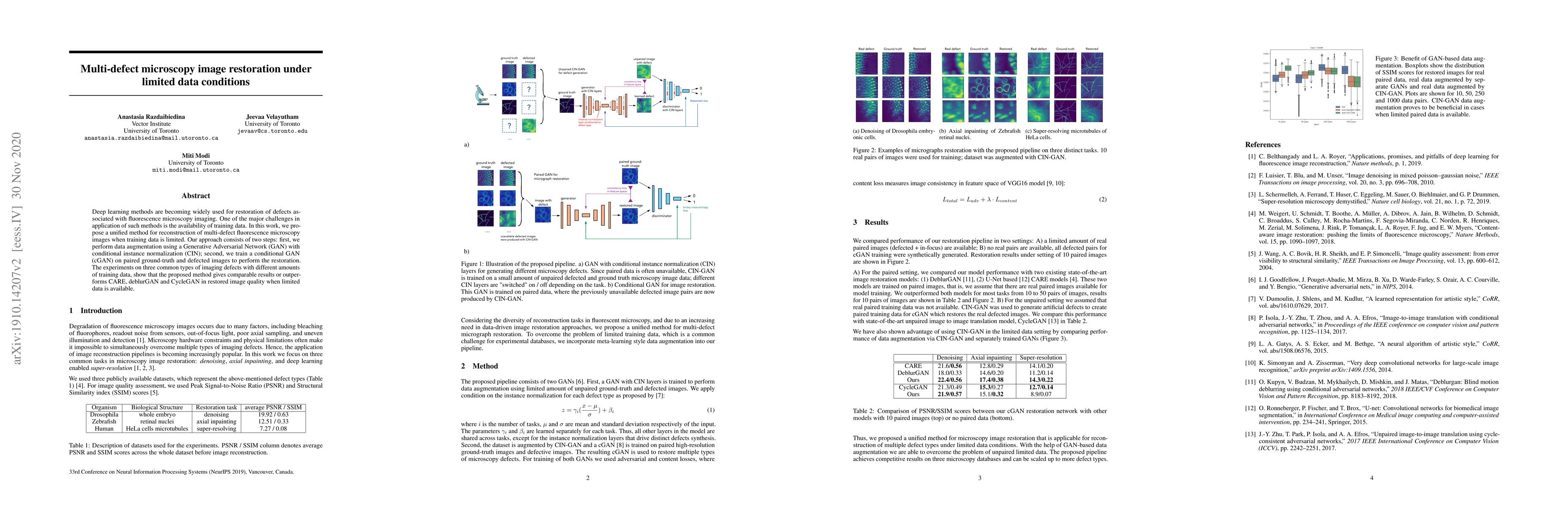
The research proposes a two-stage method for restoring multi-defect fluorescence microscopy images using limited training data. The first stage involves data augmentation with a Generative Adversarial Network (GAN) incorporating conditional instance normalization (CIN). The second stage trains a conditional GAN (cGAN) on paired ground-truth and defected images for restoration.
Discussion 0