Multi-Feature Vision Transformer via Self-Supervised Representation Learning for Improvement of COVID-19 Diagnosis
Publication
Metrics
AI Quick Summary
This paper proposes a self-supervised Vision Transformer model for diagnosing COVID-19 from chest X-ray images, utilizing both original and enhanced local phase images to improve diagnostic performance. The approach achieves 91.10% accuracy with only 10% labeled data, outperforming existing methods.
Paper Preview
Abstract
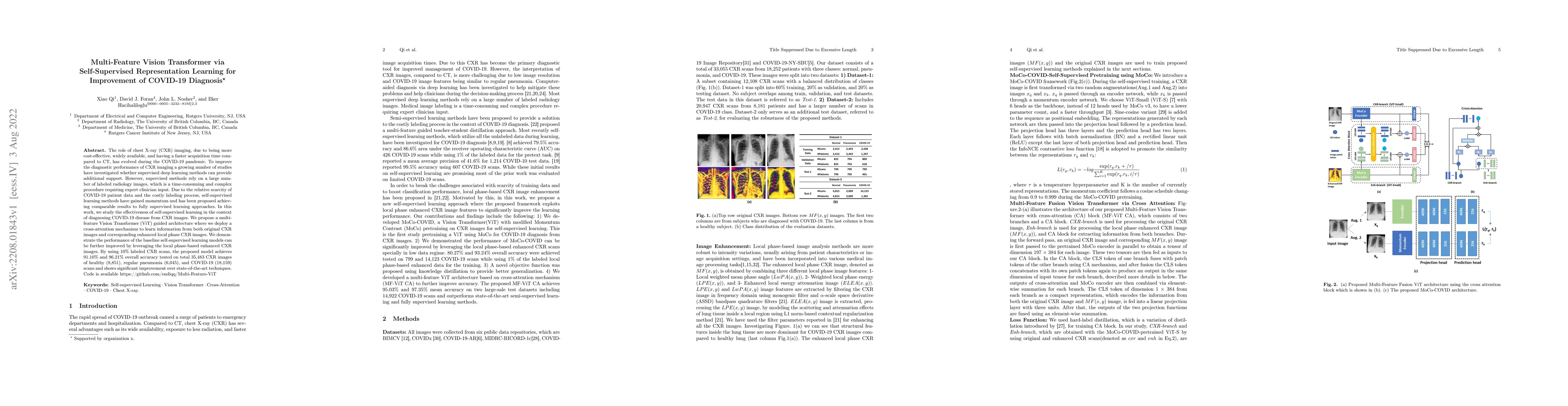
The role of chest X-ray (CXR) imaging, due to being more cost-effective, widely available, and having a faster acquisition time compared to CT, has evolved during the COVID-19 pandemic. To improve the diagnostic performance of CXR imaging a growing number of studies have investigated whether supervised deep learning methods can provide additional support. However, supervised methods rely on a large number of labeled radiology images, which is a time-consuming and complex procedure requiring expert clinician input. Due to the relative scarcity of COVID-19 patient data and the costly labeling process, self-supervised learning methods have gained momentum and has been proposed achieving comparable results to fully supervised learning approaches. In this work, we study the effectiveness of self-supervised learning in the context of diagnosing COVID-19 disease from CXR images. We propose a multi-feature Vision Transformer (ViT) guided architecture where we deploy a cross-attention mechanism to learn information from both original CXR images and corresponding enhanced local phase CXR images. We demonstrate the performance of the baseline self-supervised learning models can be further improved by leveraging the local phase-based enhanced CXR images. By using 10\% labeled CXR scans, the proposed model achieves 91.10\% and 96.21\% overall accuracy tested on total 35,483 CXR images of healthy (8,851), regular pneumonia (6,045), and COVID-19 (18,159) scans and shows significant improvement over state-of-the-art techniques. Code is available https://github.com/endiqq/Multi-Feature-ViT
AI Key Findings
Get AI-generated insights about this paper's methodology, results, significance, and more — seven facets brought into focus.
Impact
Paper Details
Authors
PDF Preview
Key Terms
Citation Network
Current paper (gray), citations (green), references (blue)
Display is limited for performance on very large graphs.
Discussion 0