Authors
Summary
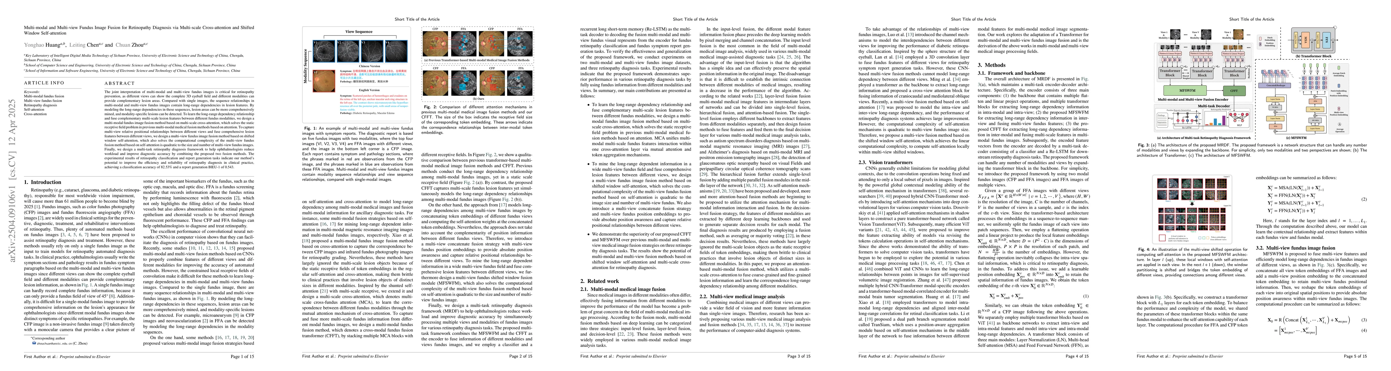
The joint interpretation of multi-modal and multi-view fundus images is critical for retinopathy prevention, as different views can show the complete 3D eyeball field and different modalities can provide complementary lesion areas. Compared with single images, the sequence relationships in multi-modal and multi-view fundus images contain long-range dependencies in lesion features. By modeling the long-range dependencies in these sequences, lesion areas can be more comprehensively mined, and modality-specific lesions can be detected. To learn the long-range dependency relationship and fuse complementary multi-scale lesion features between different fundus modalities, we design a multi-modal fundus image fusion method based on multi-scale cross-attention, which solves the static receptive field problem in previous multi-modal medical fusion methods based on attention. To capture multi-view relative positional relationships between different views and fuse comprehensive lesion features between different views, we design a multi-view fundus image fusion method based on shifted window self-attention, which also solves the computational complexity of the multi-view fundus fusion method based on self-attention is quadratic to the size and number of multi-view fundus images. Finally, we design a multi-task retinopathy diagnosis framework to help ophthalmologists reduce workload and improve diagnostic accuracy by combining the proposed two fusion methods. The experimental results of retinopathy classification and report generation tasks indicate our method's potential to improve the efficiency and reliability of retinopathy diagnosis in clinical practice, achieving a classification accuracy of 82.53\% and a report generation BlEU-1 of 0.543.
AI Key Findings
Generated Jun 09, 2025
Methodology
The paper proposes a multi-modal and multi-view fundus image fusion framework for retinopathy diagnosis using multi-scale cross-attention and shifted window self-attention mechanisms. It combines multi-view fundus image fusion (MFSWFM) and multi-modal fundus image fusion (CFFT) methods to capture comprehensive lesion features from different modalities and views.
Key Results
- The proposed method achieved a classification accuracy of 82.53% on the SFPD dataset.
- The model outperformed other multi-modal and multi-view fusion methods in both single-label retinopathy classification and multi-label retinopathy classification tasks on the MSRD dataset.
- The report generation task using BLEU-1, BLEU-2, BLEU-3, BLEU-4, METEOR, ROUGE-R, and CIDEr metrics showed that the proposed method surpassed existing methods by significant margins.
Significance
This research is significant as it enhances retinopathy diagnosis efficiency and reliability in clinical practice by effectively fusing multi-modal and multi-view fundus images, which can assist ophthalmologists in analyzing complex fundus images and generating accurate symptom reports.
Technical Contribution
The paper introduces a novel multi-task framework that simultaneously processes fundus images of different views and modalities for retinopathy pathological classification and symptom report generation tasks, leveraging transformer blocks for scalability.
Novelty
The proposed method distinguishes itself by employing shifted window self-attention to address computational complexity in multi-view fundus image fusion, and multi-scale cross-attention for multi-modal fundus image fusion, outperforming existing methods in both classification and report generation tasks.
Limitations
- The study relied on private datasets, limiting the generalizability of results to publicly available datasets.
- Computational complexity and model parameters might still be considerable for some applications.
Future Work
- Explore the applicability of the proposed method on other 3D multi-modal medical imaging datasets.
- Investigate methods to reduce model complexity and inference time without compromising performance.
Paper Details
PDF Preview
Citation Network
Current paper (gray), citations (green), references (blue)
Display is limited for performance on very large graphs.
Similar Papers
Found 4 papersMulti-Modal Brain Tumor Segmentation via 3D Multi-Scale Self-attention and Cross-attention
Chuan Zhou, Yonghao Huang, Leiting Chen
Cross-Fundus Transformer for Multi-modal Diabetic Retinopathy Grading with Cataract
Yi Xu, Rui Feng, Fan Xiao et al.
DMS-Net:Dual-Modal Multi-Scale Siamese Network for Binocular Fundus Image Classification
Hao Tang, Zitong Wang, Ruiting Dai et al.
Multi-scale Quaternion CNN and BiGRU with Cross Self-attention Feature Fusion for Fault Diagnosis of Bearing
Yan Li, Shenghong Luo, Huanbai Liu et al.
No citations found for this paper.
Comments (0)