Multi-modal Brain Tumor Segmentation via Missing Modality Synthesis and Modality-level Attention Fusion
Publication
Metrics
AI Quick Summary
This paper proposes a framework called Modality-Level Attention Fusion Network (MAF-Net) to synthesize unavailable T1ce MR modalities from existing T1, T2, and FLAIR images, enhancing brain tumor segmentation. The MAF-Net uses patchwise contrastive learning and modality-level attention fusion to achieve superior synthesis and segmentation performance.
Paper Preview
Abstract
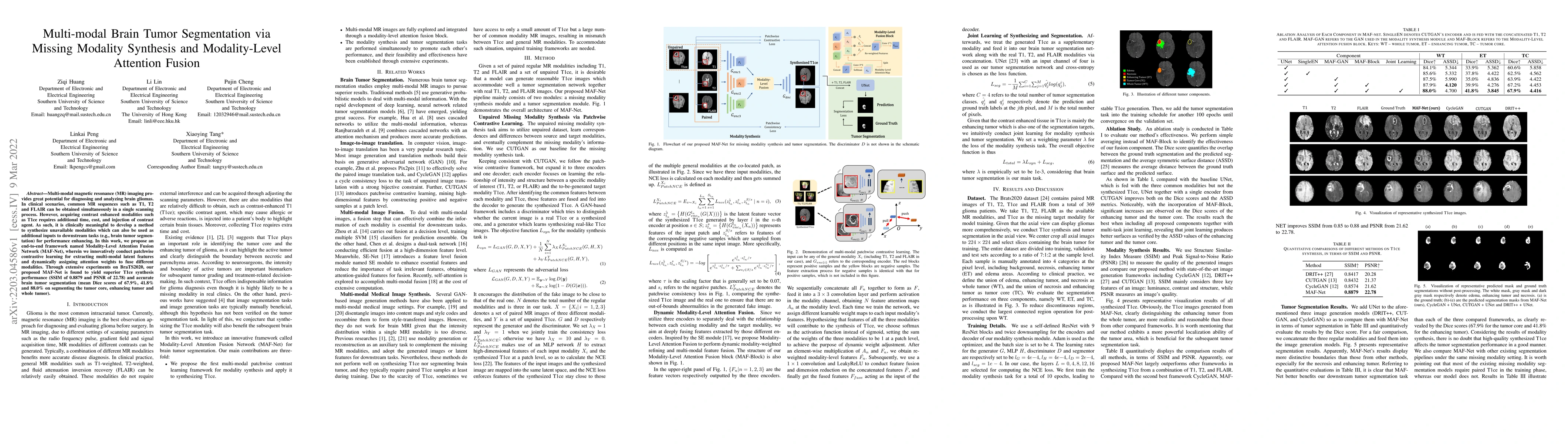
Multi-modal magnetic resonance (MR) imaging provides great potential for diagnosing and analyzing brain gliomas. In clinical scenarios, common MR sequences such as T1, T2 and FLAIR can be obtained simultaneously in a single scanning process. However, acquiring contrast enhanced modalities such as T1ce requires additional time, cost, and injection of contrast agent. As such, it is clinically meaningful to develop a method to synthesize unavailable modalities which can also be used as additional inputs to downstream tasks (e.g., brain tumor segmentation) for performance enhancing. In this work, we propose an end-to-end framework named Modality-Level Attention Fusion Network (MAF-Net), wherein we innovatively conduct patchwise contrastive learning for extracting multi-modal latent features and dynamically assigning attention weights to fuse different modalities. Through extensive experiments on BraTS2020, our proposed MAF-Net is found to yield superior T1ce synthesis performance (SSIM of 0.8879 and PSNR of 22.78) and accurate brain tumor segmentation (mean Dice scores of 67.9%, 41.8% and 88.0% on segmenting the tumor core, enhancing tumor and whole tumor).
AI Key Findings
Get AI-generated insights about this paper's methodology, results, significance, and more — seven facets brought into focus.
Impact
Paper Details
Authors
PDF Preview
Key Terms
Citation Network
Current paper (gray), citations (green), references (blue)
Display is limited for performance on very large graphs.
Discussion 0