The study developed an automated pipeline integrating multi-modal co-registration, deep neural networks for liver segmentation, and STAD features for fibrosis staging. The pipeline was trained on multi-centre, multi-modal MRI data with limited annotations, using 3D nnUNet for segmentation and Random Forest classifiers for fibrosis staging.
Multi-modal Liver Segmentation and Fibrosis Staging Using Real-world MRI Images
Publication
Metrics
Quick Answers
What methodology did the authors use?
The study developed an automated pipeline integrating multi-modal co-registration, deep neural networks for liver segmentation, and STAD features for fibrosis staging. The pipeline was trained on multi-centre, multi-modal MRI data with limited annotations, using 3D nnUNet for segmentation and Random Forest classifiers for fibrosis staging. More in Methodology →
What are the key results?
The pipeline achieved top-tier performance across all competition subtasks with excellent generalisability to different MRI modalities. — Model-All-Norm demonstrated improved Dice scores and smaller Hausdorff distances on out-of-distribution data compared to in-distribution data. More in Key Results →
Why is this work significant?
This research provides a rapid, reproducible framework for non-invasive liver fibrosis assessment, supporting early diagnosis and clinical decision-making. It addresses the limitations of invasive biopsies and enables frequent monitoring of disease progression. More in Significance →
What are the main limitations?
Inaccurate pseudo-labels from co-registration introduced bias during segmentation training, leading to potential overfitting. — Feature selection was limited by reliance on handcrafted features, which may underperform compared to deep learning approaches. More in Limitations →
Abstract
Liver fibrosis represents the accumulation of excessive extracellular matrix caused by sustained hepatic injury. It disrupts normal lobular architecture and function, increasing the chances of cirrhosis and liver failure. Precise staging of fibrosis for early diagnosis and intervention is often invasive, which carries risks and complications. To address this challenge, recent advances in artificial intelligence-based liver segmentation and fibrosis staging offer a non-invasive alternative. As a result, the CARE 2025 Challenge aimed for automated methods to quantify and analyse liver fibrosis in real-world scenarios, using multi-centre, multi-modal, and multi-phase MRI data. This challenge included tasks of precise liver segmentation (LiSeg) and fibrosis staging (LiFS). In this study, we developed an automated pipeline for both tasks across all the provided MRI modalities. This pipeline integrates pseudo-labelling based on multi-modal co-registration, liver segmentation using deep neural networks, and liver fibrosis staging based on shape, textural, appearance, and directional (STAD) features derived from segmentation masks and MRI images. By solely using the released data with limited annotations, our proposed pipeline demonstrated excellent generalisability for all MRI modalities, achieving top-tier performance across all competition subtasks. This approach provides a rapid and reproducible framework for quantitative MRI-based liver fibrosis assessment, supporting early diagnosis and clinical decision-making. Code is available at https://github.com/YangForever/care2025_liver_biodreamer.
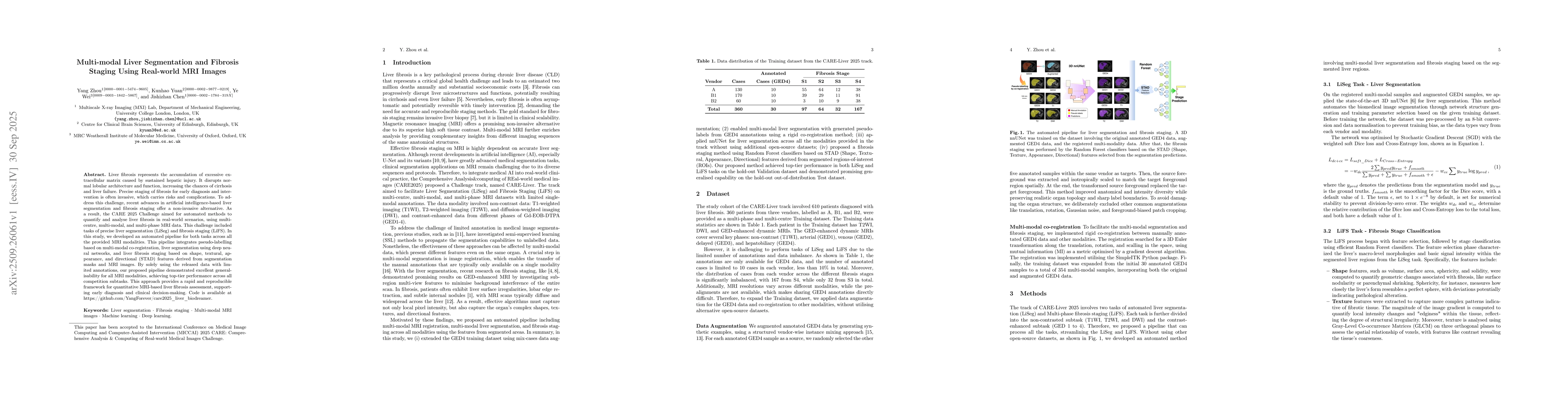
Key Findings, in focus
Seven facets of this paper, analysed and brought into focus by AI.
This research provides a rapid, reproducible framework for non-invasive liver fibrosis assessment, supporting early diagnosis and clinical decision-making.
- The pipeline achieved top-tier performance across all competition subtasks with excellent generalisability to different MRI modalities.
- Model-All-Norm demonstrated improved Dice scores and smaller Hausdorff distances on out-of-distribution data compared to in-distribution data.
- The feature importance analysis showed shape-based and directional features were most significant for fibrosis detection, with directional features like coherence and anisotropy showing high importance.
This research provides a rapid, reproducible framework for non-invasive liver fibrosis assessment, supporting early diagnosis and clinical decision-making. It addresses the limitations of invasive biopsies and enables frequent monitoring of disease progression.
The integration of multi-modal co-registration, 3D nnUNet for segmentation, and STAD features for fibrosis staging represents a novel technical contribution for automated liver fibrosis analysis.
The work's novelty lies in its use of real-world, multi-centre, multi-modal MRI data for automated liver segmentation and fibrosis staging without requiring additional large open-source datasets, providing a clinically interpretable framework for fibrosis assessment.
- Inaccurate pseudo-labels from co-registration introduced bias during segmentation training, leading to potential overfitting.
- Feature selection was limited by reliance on handcrafted features, which may underperform compared to deep learning approaches.
- Investigate non-rigid co-registration methods to improve registration accuracy and downstream task performance.
- Explore neural network-based methods for better generalization, especially with larger annotated datasets.
- Incorporate more shape-based and directional features while reducing intensity-based appearance features in training.
Discussion 0