Multi-Resolution Histopathology Patch Graphs for Ovarian Cancer Subtyping
Publication
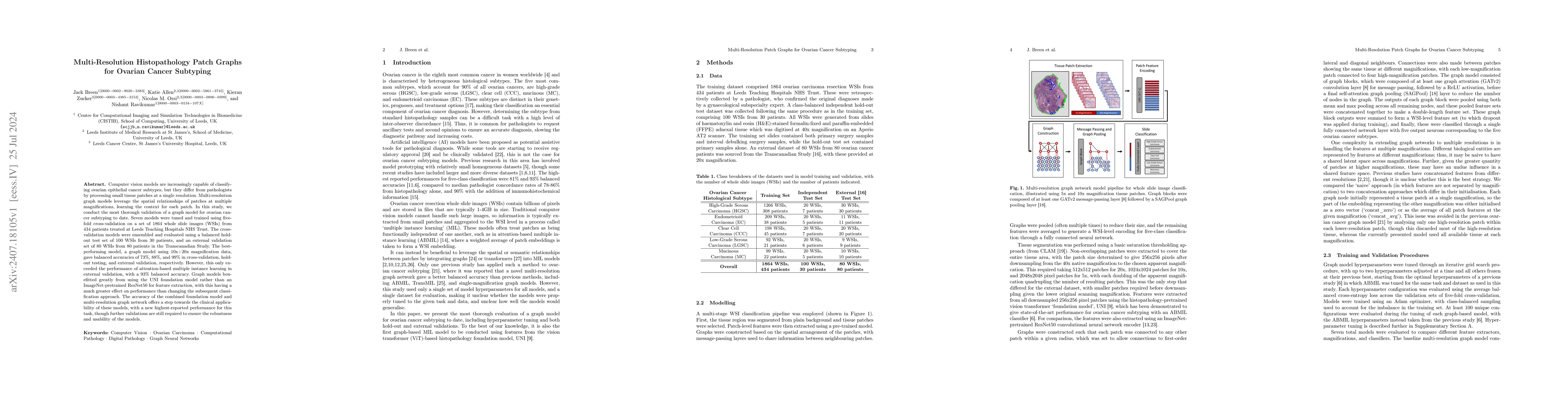
Metrics
AI Quick Summary
This study introduces multi-resolution graph models to improve ovarian cancer subtyping, achieving 99% balanced accuracy in external validation using a foundation model. Despite surpassing other methods in external settings, further validations are necessary to ensure model robustness.
Paper Preview
Abstract
Computer vision models are increasingly capable of classifying ovarian epithelial cancer subtypes, but they differ from pathologists by processing small tissue patches at a single resolution. Multi-resolution graph models leverage the spatial relationships of patches at multiple magnifications, learning the context for each patch. In this study, we conduct the most thorough validation of a graph model for ovarian cancer subtyping to date. Seven models were tuned and trained using five-fold cross-validation on a set of 1864 whole slide images (WSIs) from 434 patients treated at Leeds Teaching Hospitals NHS Trust. The cross-validation models were ensembled and evaluated using a balanced hold-out test set of 100 WSIs from 30 patients, and an external validation set of 80 WSIs from 80 patients in the Transcanadian Study. The best-performing model, a graph model using 10x+20x magnification data, gave balanced accuracies of 73%, 88%, and 99% in cross-validation, hold-out testing, and external validation, respectively. However, this only exceeded the performance of attention-based multiple instance learning in external validation, with a 93% balanced accuracy. Graph models benefitted greatly from using the UNI foundation model rather than an ImageNet-pretrained ResNet50 for feature extraction, with this having a much greater effect on performance than changing the subsequent classification approach. The accuracy of the combined foundation model and multi-resolution graph network offers a step towards the clinical applicability of these models, with a new highest-reported performance for this task, though further validations are still required to ensure the robustness and usability of the models.
AI Key Findings
Get AI-generated insights about this paper's methodology, results, significance, and more — seven facets brought into focus.
Impact
Paper Details
Authors
PDF Preview
Key Terms
Citation Network
Current paper (gray), citations (green), references (blue)
Display is limited for performance on very large graphs.
Discussion 0