Publication
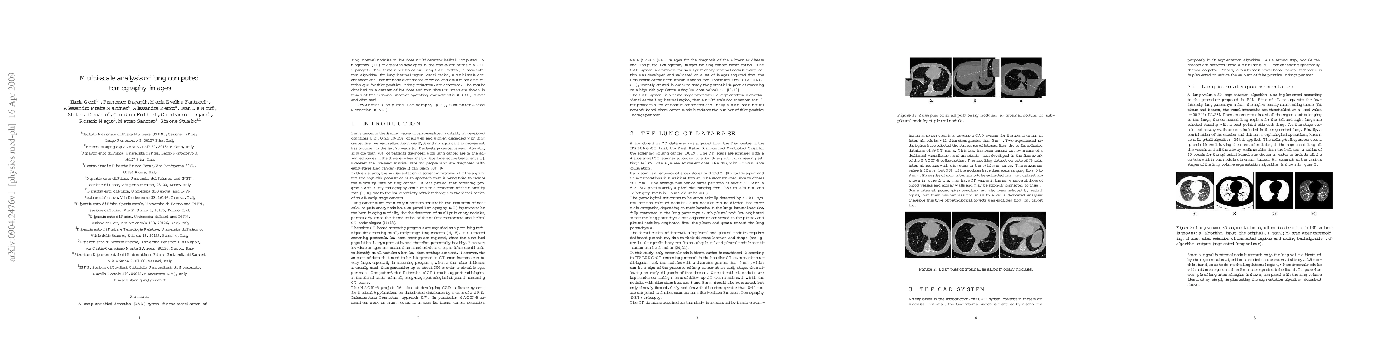
Metrics
AI Quick Summary
This paper presents a CAD system for detecting lung nodules in low-dose CT scans, comprising segmentation, nodule candidate selection, and false positive reduction modules. The system's performance is evaluated using FROC curves on a dataset of low-dose, thin-slice CT scans.
Paper Preview
Abstract
A computer-aided detection (CAD) system for the identification of lung internal nodules in low-dose multi-detector helical Computed Tomography (CT) images was developed in the framework of the MAGIC-5 project. The three modules of our lung CAD system, a segmentation algorithm for lung internal region identification, a multi-scale dot-enhancement filter for nodule candidate selection and a multi-scale neural technique for false positive finding reduction, are described. The results obtained on a dataset of low-dose and thin-slice CT scans are shown in terms of free response receiver operating characteristic (FROC) curves and discussed.
AI Key Findings
Get AI-generated insights about this paper's methodology, results, significance, and more — seven facets brought into focus.
Impact
Paper Details
PDF Preview
Key Terms
Citation Network
Current paper (gray), citations (green), references (blue)
Display is limited for performance on very large graphs.
Discussion 0