This paper proposes an advanced segmentation framework for the left atrium from cardiac MRI using a multi-scale feature fusion approach that combines DINOv2 with a UNet-style decoder, enhancing spatial resolution and accuracy. The method achieves superior performance with a 92.3% Dice and 84.1% IoU score, demonstrating its effectiveness over existing baseline models.
Accurate segmentation of the left atrium (LA) from late gadolinium-enhanced magnetic resonance imaging plays a vital role in visualizing diseased atrial structures, enabling the diagnosis and management of cardiovascular diseases. It is particularly essential for planning treatment with ablation therapy, a key intervention for atrial fibrillation (AF). However, manual segmentation is time-intensive and prone to inter-observer variability, underscoring the need for automated solutions. Class-agnostic foundation models like DINOv2 have demonstrated remarkable feature extraction capabilities in vision tasks. However, their lack of domain specificity and task-specific adaptation can reduce spatial resolution during feature extraction, impacting the capture of fine anatomical detail in medical imaging. To address this limitation, we propose a segmentation framework that integrates DINOv2 as an encoder with a UNet-style decoder, incorporating multi-scale feature fusion and input image integration to enhance segmentation accuracy. The learnable weighting mechanism dynamically prioritizes hierarchical features from different encoder blocks of the foundation model, optimizing feature selection for task relevance. Additionally, the input image is reintroduced during the decoding stage to preserve high-resolution spatial details, addressing limitations of downsampling in the encoder. We validate our approach on the LAScarQS 2022 dataset and demonstrate improved performance with a 92.3% Dice and 84.1% IoU score for giant architecture compared to the nnUNet baseline model. These findings emphasize the efficacy of our approach in advancing the field of automated left atrium segmentation from cardiac MRI.
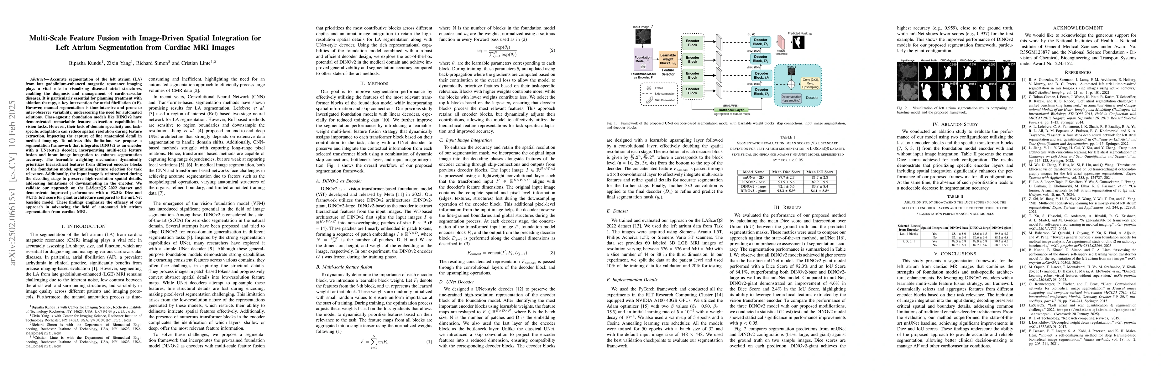
Generated Jun 11, 2025
The research proposes a segmentation framework that integrates DINOv2 as an encoder with a UNet-style decoder, incorporating multi-scale feature fusion and input image integration to enhance segmentation accuracy. The framework dynamically prioritizes hierarchical features from different encoder blocks and reintroduces the input image during decoding to preserve high-resolution spatial details.
This research is significant as it addresses the challenge of accurate left atrium segmentation from cardiac MRI images, which is crucial for diagnosing and managing cardiovascular diseases, especially atrial fibrillation. The proposed method improves upon existing automated segmentation solutions by effectively leveraging hierarchical features extracted by vision transformer encoders.
The main technical contribution is a segmentation framework that combines DINOv2 as a feature extractor with a UNet-style decoder, incorporating multi-scale feature fusion and input image integration to improve segmentation accuracy in medical imaging.
This work is novel in its approach to adapt a class-agnostic foundation model (DINOv2) for medical image segmentation by integrating it with a task-specific UNet-style decoder, dynamically prioritizing relevant encoder blocks, and reintroducing the input image during decoding to maintain high-resolution spatial details.
Current paper (gray), citations (green), references (blue)
Display is limited for performance on very large graphs.
Marta Varela, Christoforos Galazis, Anil A. Bharath et al.
Gege Ma, Wentao Zhu, Duo Zhang et al.
No citations found for this paper.
Comments (0)