Summary
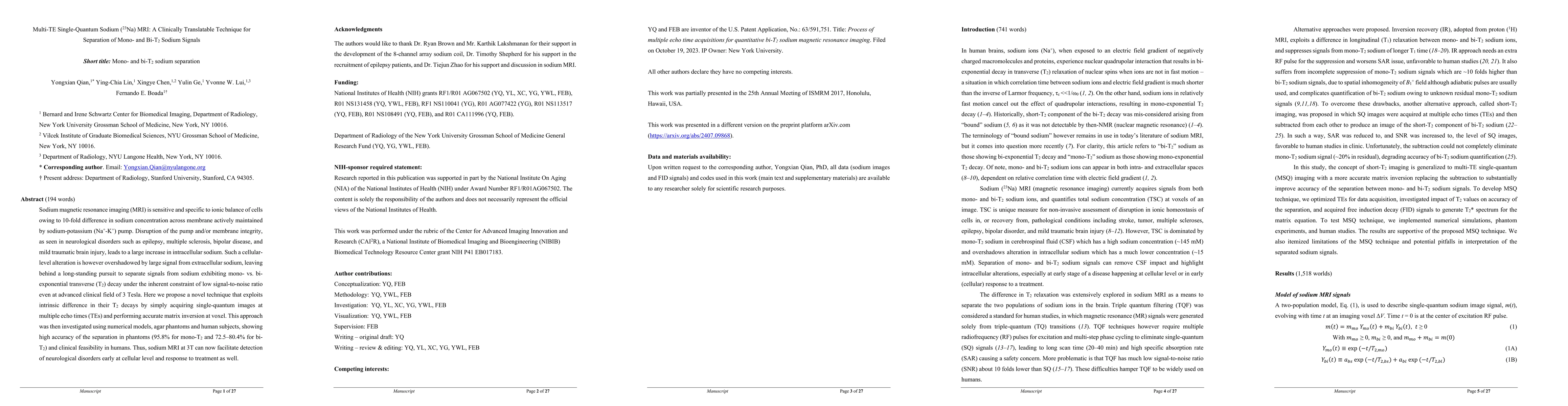
Sodium magnetic resonance imaging (MRI) is sensitive and specific to ionic balance of cells owing to 10 fold difference in sodium concentration across membrane actively maintained by sodium potassium (Na+ K+) pump. Disruption of the pump and membrane integrity, as seen in neurological disorders such as epilepsy, multiple sclerosis, bipolar disease, and mild traumatic brain injury, leads to a large increase in intracellular sodium. Such a cellular level alteration is however overshadowed by large signal from extracellular sodium, leaving behind a long standing pursuit to separate signals from sodium exhibiting mono vs biexponential transverse (T2) decay under the inherent constraint of low signal to noise ratio even at advanced clinical field of 3 Tesla. Here we propose a novel technique that exploits intrinsic difference in their T2 decays by simply acquiring single quantum images at multiple echo times (TEs) and performing accurate matrix inversion at voxel. This approach was then investigated using numerical models, agar phantoms and human subjects, showing high accuracy of the separation in phantoms (95.8 percent for monoT2 and 72.5 to 80.4 percent for biT2) and clinical feasibility in humans. Thus, sodium MRI at 3T can now facilitate detection of neurological disorders early at cellular level and response to treatment as well. Keywords. sodium MRI, single quantum MRI, triple quantum MRI, neuroimaging, neurodegeneration
AI Key Findings
Get AI-generated insights about this paper's methodology, results, and significance.
Paper Details
PDF Preview
Citation Network
Current paper (gray), citations (green), references (blue)
Display is limited for performance on very large graphs.
Similar Papers
Found 4 papersAnalysis of blurring due to short T2 decay at different resolutions in 23Na MRI
Zidan Yu, Olga Dergachyova, Shota Hodono et al.
Rician Denoising Diffusion Probabilistic Models For Sodium Breast MRI Enhancement
Shuaiyu Yuan, Tristan Whitmarsh, Dimitri A Kessler et al.
Compartment-specific estimation of T2 and T2* with diffusion-PEPTIDE MRI
Hui Zhang, Ting Gong, Kawin Setsompop et al.
No citations found for this paper.
Comments (0)