Multifractality in Surface Potential for Cancer Diagnosis
Publication
Metrics
AI Quick Summary
This paper demonstrates the use of multifractal analysis for differentiating between normal and cancerous cells through surface potential imaging using Kelvin probe force microscopy. The study shows that adaptive threshold binarization improves cancer diagnosis compared to median threshold, suggesting multifractality as a promising new biomarker for cancer detection.
Paper Preview
Abstract
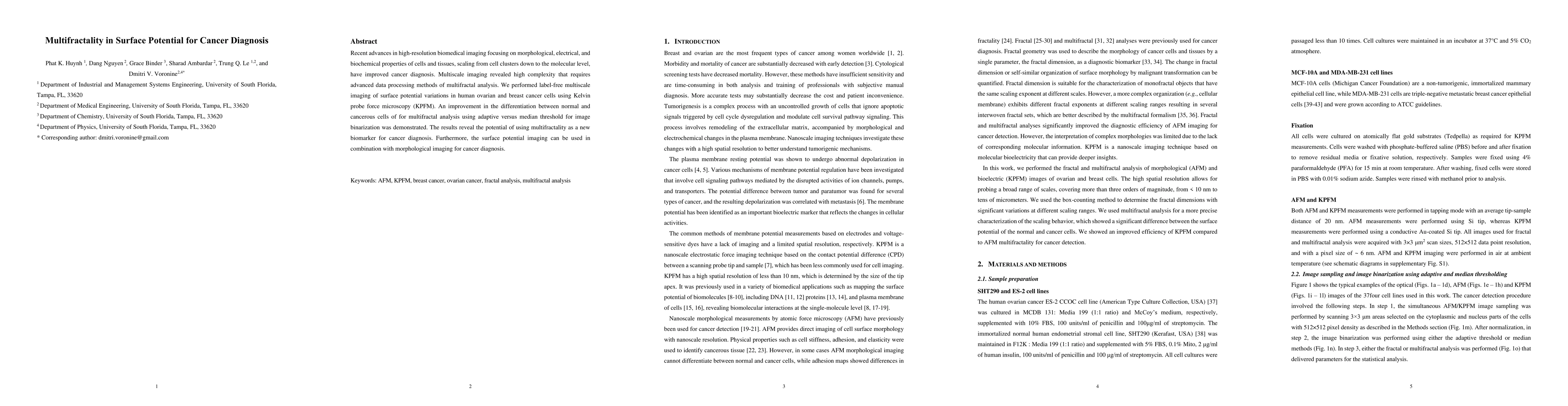
Recent advances in high-resolution biomedical imaging focusing on morphological, electrical, and biochemical properties of cells and tissues, scaling from cell clusters down to the molecular level, have improved cancer diagnosis. Multiscale imaging revealed high complexity that requires advanced data processing methods of multifractal analysis. We performed label-free multiscale imaging of surface potential variations in human ovarian and breast cancer cells using Kelvin probe force microscopy (KPFM). An improvement in the differentiation between normal and cancerous cells of for multifractal analysis using adaptive versus median threshold for image binarization was demonstrated. The results reveal the potential of using multifractality as a new biomarker for cancer diagnosis. Furthermore, the surface potential imaging can be used in combination with morphological imaging for cancer diagnosis.
AI Key Findings
Get AI-generated insights about this paper's methodology, results, significance, and more — seven facets brought into focus.
Impact
Paper Details
Authors
PDF Preview
Key Terms
Citation Network
Current paper (gray), citations (green), references (blue)
Display is limited for performance on very large graphs.
Discussion 0