Quick Summary:

This paper introduces a multimarginal Wasserstein barycenter method for normalizing and augmenting hematoxylin and eosin (H&E) stained images, addressing variations due to lab protocols and scanners. The approach effectively drives stain normalization and augmentation using additional reference images, achieving superior results compared to existing methods and state-of-the-art performance in nuclei segmentation tasks.

Summary

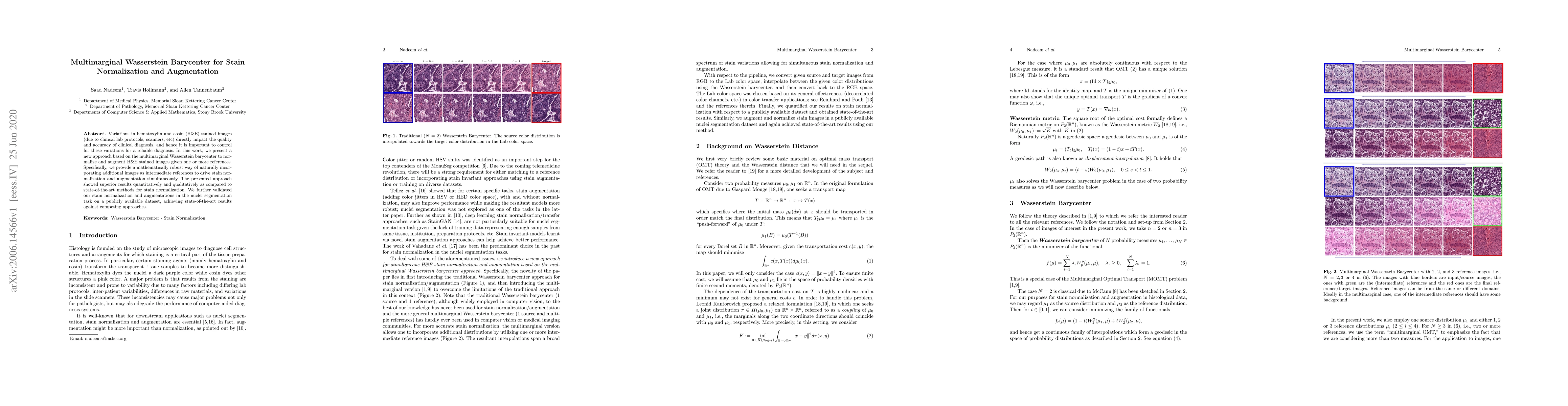
Variations in hematoxylin and eosin (H&E) stained images (due to clinical lab protocols, scanners, etc) directly impact the quality and accuracy of clinical diagnosis, and hence it is important to control for these variations for a reliable diagnosis. In this work, we present a new approach based on the multimarginal Wasserstein barycenter to normalize and augment H&E stained images given one or more references. Specifically, we provide a mathematically robust way of naturally incorporating additional images as intermediate references to drive stain normalization and augmentation simultaneously. The presented approach showed superior results quantitatively and qualitatively as compared to state-of-the-art methods for stain normalization. We further validated our stain normalization and augmentations in the nuclei segmentation task on a publicly available dataset, achieving state-of-the-art results against competing approaches.

AI Key Findings

Get AI-generated insights about this paper's methodology, results, and significance.

Paper Details

PDF Preview

Download PDF

Key Terms

normalization (0.307)
diagnosis (0.252)
clinical (0.238)
images (0.208)
variations (0.198)
state art (0.154)
art (0.153)
compared state (0.151)
competing (0.149)
validated (0.144)
qualitatively (0.143)
publicly available (0.142)
art methods (0.142)
augmentation (0.141)
new approach (0.139)
work present (0.137)
quantitatively (0.137)
approach based (0.137)
drive (0.137)
publicly (0.133)

Citation Network

Current paper (gray), citations (green), references (blue)

Current Paper
Citations
References
Click to view
Title Authors Year Actions
StainSWIN: Vision transformer-based stain normalization for histopathology image analysis
Elif Baykal Kablan, Selen Ayas
2024
View Paper
Stain SAN: simultaneous augmentation and normalization for histopathology images.
Taebin Kim, Yao Li, Benjamin C. Calhoun, A. Thennavan, L. Carey, W. Symmans, M. A. Troester, C. Perou, J. S. Marron
2024
View Paper
Computational pathology: A survey review and the way forward
M. S. Hosseini, B. Bejnordi, Vincent Quoc-Huy Trinh, Lyndon Chan, Danial Hasan, Xingwen Li, Stephen Yang, Taehyo Kim, Haochen Zhang, Theodore Wu, Kajanan Chinniah, Sina Maghsoudlou, Ryan Zhang, Jiadai Zhu, Samir Khaki, Andrei Buin, Fatemeh Chaji, Ala Salehi, Bich Ngoc Nguyen, Dimitris Samaras, K. Plataniotis
2024
View Paper
Stain normalization methods for histopathology image analysis: A comprehensive review and experimental comparison
Md. Ziaul Hoque, A. Keskinarkaus, Pia Nyberg, T. Seppänen
2023
View Paper
Regional realness-aware generative adversarial networks for stain normalization
Elif Baykal Kablan
2023
View Paper
Multimodal deep learning to predict prognosis in adult and pediatric brain tumors
S. Steyaert, Y. Qiu, Yuanning Zheng, P. Mukherjee, Hannes Vogel, O. Gevaert
2023
View Paper
Stain-invariant self supervised learning for histopathology image analysis
Alexandre Tiard, A. Wong, D. J. Ho, Yang Wu, Eliram Nof, Stefano Soatto, S. Nadeem
2022
View Paper
Multimodal data fusion of adult and pediatric brain tumors with deep learning
S. Steyaert, Y. Qiu, Y. Zheng, P. Mukherjee, H. Vogel, O. Gevaert
2022
View Paper
Stain-Adaptive Self-Supervised Learning for Histopathology Image Analysis
Haili Ye, Da-han Wang
2022
View Paper
Colour adaptive generative networks for stain normalisation of histopathology images
Cong Cong, Sidong Liu, A. Ieva, M. Pagnucco, S. Berkovsky, Yang Song
2022
View Paper
RandStainNA: Learning Stain-Agnostic Features from Histology Slides by Bridging Stain Augmentation and Normalization
Yiqing Shen, Yulin Luo, Dinggang Shen, Jing Ke
2022
View Paper
MixPatch: A New Method for Training Histopathology Image Classifiers
Youngjin Park, Mujin Kim, Murtaza Ashraf, Y. Ko, M. Y. Yi
2022
View Paper
ALLWAS: Active Learning on Language models in WASserstein space
Anson Bastos, Manohar Kaul
2021
View Paper
Structure-Preserving Multi-Domain Stain Color Augmentation using Style-Transfer with Disentangled Representations
S. Wagner, Nadieh Khalili, Raghav Sharma, M. Boxberg, C. Marr, Walter de Back, Tingying Peng
2021
View Paper
LAMBO: Landmarks Augmentation With Manifold-Barycentric Oversampling
Iaroslav Bespalov, N. Buzun, Oleg Kachan, D. Dylov
2021
View Paper
Evaluating Registrations of Serial Sections With Distortions of the Ground Truths
Oleg Lobachev, Takuya Funatomi, Alexander Pfaffenroth, R. Förster, Lars Knudsen, C. Wrede, M. Guthe, D. Haberthür, R. Hlushchuk, T. Salaets, J. Toelen, Simone Gaffling, C. Mühlfeld, R. Grothausmann
2020
View Paper
Distributed Optimization with Quantization for Computing Wasserstein Barycenters
R. Krawtschenko, César A. Uribe, A. Gasnikov, P. Dvurechensky
2020
View Paper
ACC-GAN: Cross Scanner Robustness With Annotation Consistency Guided Cycle-GAN
Chien-Yu Chiou, Hung-Wen Tsai, Wei-Jong Yang, Chih-Hsien Lee, Ming Ting Sun, Maria Gabrani, Kuo-Sheng Cheng, Meng-Ling Wu, Pau-Choo Chung
2024
View Paper
Semi-supervised Adversarial Learning for Stain Normalisation in Histopathology Images
Cong Cong, Sidong Liu, A. Ieva, M. Pagnucco, S. Berkovsky, Yang Song
2021
View Paper
Stain Mix-Up: Unsupervised Domain Generalization for Histopathology Images
Jia-Ren Chang, Min-Sheng Wu, Wei-Hsiang Yu, Chi-Chung Chen, Cheng-Kung Yang, Yen-Yu Lin, Chao-Yuan Yeh
2021
View Paper
Contrastive Learning Based Stain Normalization Across Multiple Tumor in Histopathology
Jing Ke, Yiqing Shen, Xiaoyao Liang, D. Shen
2021
View Paper

Comments (0)

This website uses cookies to ensure you get the best experience. Learn more