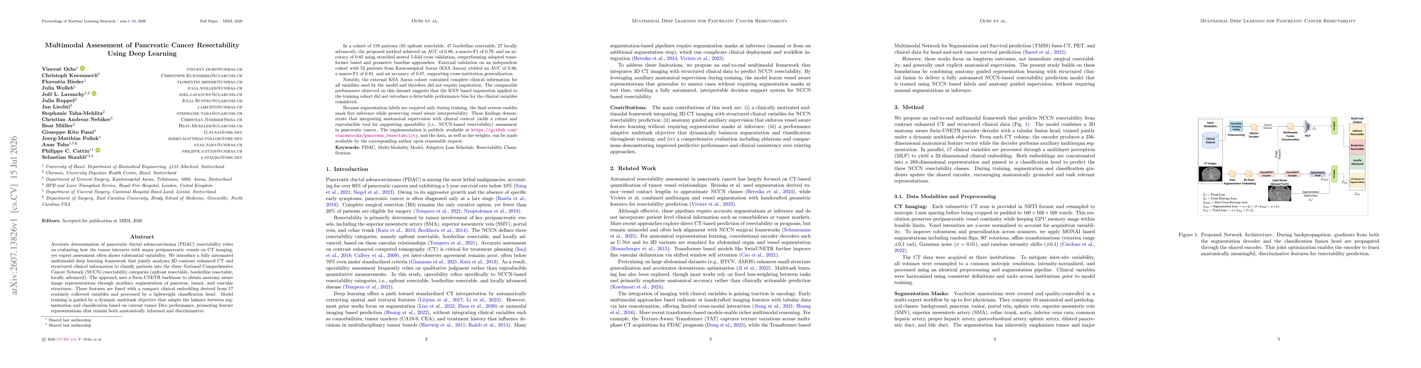
A fully automated multimodal deep learning framework that jointly analyzes 3D contrast-enhanced CT imaging and structured clinical information to classify PDAC resectability into NCCN categories. It uses a Swin-UNETR backbone to derive anatomy-aware representations via auxiliary segmentation of pancreas, tumor, and vascular structures, then fuses these features with a compact clinical embedding from 17 variables. Training employs a dynamic multitask objective balancing segmentation and classification based on current tumour Dice performance to promote anatomically informed and discriminative representations.
Multimodal Assessment of Pancreatic Cancer Resectability Using Deep Learning
Publication
Metrics
AI Quick Summary
A fully automated deep learning framework integrates 3D CT images and 17 clinical variables to classify pancreatic cancer resectability into upfront resectable, borderline, or locally advanced. Using a Swin-UNETR backbone for anatomy-aware segmentation and a dynamic multitask objective, the model fuses imaging features with clinical data to improve consistent, expert-level assessment for surgical planning.
Quick Answers
What is "Multimodal Assessment of Pancreatic Cancer Resectability Using Deep Learning" about?
A fully automated deep learning framework integrates 3D CT images and 17 clinical variables to classify pancreatic cancer resectability into upfront resectable, borderline, or locally advanced. Using a Swin-UNETR backbone for anatomy-aware segmentation and a dynamic multitask objective, the model fuses imaging features with clinical data to improve consistent, expert-level assessment for surgical planning.
What methodology did the authors use?
A fully automated multimodal deep learning framework that jointly analyzes 3D contrast-enhanced CT imaging and structured clinical information to classify PDAC resectability into NCCN categories. It uses a Swin-UNETR backbone to derive anatomy-aware representations via auxiliary segmentation of pancreas, tumor, and vascular structures, then fuses these features with a compact clinical embedding from 17 variables. Training employs a dynamic multitask... More in Methodology →
What are the key results?
In a cohort of 159 patients, the model achieved AUC 0.86, macro-F1 0.79, and accuracy 0.85 with stratified nested 5-fold cross-validation. — External validation on 52 patients from KSAAarau yielded AUC 0.86, macro-F1 0.81, and accuracy 0.87, supporting cross-institution generalization. More in Key Results →
Why is this work significant?
The study demonstrates that integrating anatomy-aware imaging features with clinical variables yields robust and reproducible resectability assessments, potentially reducing inter-observer variability in NCCN-based decision-making for PDAC and enabling cross-institution generalization without imputation biases. More in Significance →
What are the main limitations?
Relatively modest external validation size may limit generalizability across broader patient populations. — Dependence on segmentation labels during training could constrain applicability in settings lacking annotated data for pancreas, tumor, and vessels. More in Limitations →
Abstract
Accurate determination of pancreatic ductal adenocarcinoma (PDAC) resectability relies on evaluating how the tumor interacts with major peripancreatic vessels on CT imaging, yet expert assessment often shows substantial variability. We introduce a fully automated multimodal deep learning framework that jointly analyzes 3D contrast enhanced CT and structured clinical information to classify patients into the three National Comprehensive Cancer Network (NCCN) resectability categories (upfront resectable, borderline resectable, locally advanced). The approach uses a Swin-UNETR backbone to obtain anatomy aware image representations through auxiliary segmentation of pancreas, tumor, and vascular structures. These features are fused with a compact clinical embedding derived from 17 routinely collected variables and processed by a lightweight classification head. Model training is guided by a dynamic multitask objective that adapts the balance between segmentation and classification based on current tumor Dice performance, promoting feature representations that remain both anatomically informed and discriminative.
Key Findings, in focus
Seven facets of this paper, analysed and brought into focus by AI.
The study demonstrates that integrating anatomy-aware imaging features with clinical variables yields robust and reproducible resectability assessments, potentially reducing inter-observer variability in NCCN-based decision-making for PDAC and enabling cross-institution generalization without imputation biases.
- In a cohort of 159 patients, the model achieved AUC 0.86, macro-F1 0.79, and accuracy 0.85 with stratified nested 5-fold cross-validation.
- External validation on 52 patients from KSAAarau yielded AUC 0.86, macro-F1 0.81, and accuracy 0.87, supporting cross-institution generalization.
- The external cohort had complete clinical information for all variables, enabling a mask-free inference regime and preserving vessel-aware interpretability.
The study demonstrates that integrating anatomy-aware imaging features with clinical variables yields robust and reproducible resectability assessments, potentially reducing inter-observer variability in NCCN-based decision-making for PDAC and enabling cross-institution generalization without imputation biases.
Introduction of a multimodal framework that fuses Swin-UNETR-based anatomy-aware CT representations with a compact clinical embedding and a dynamic multitask loss to balance segmentation and classification objectives for NCCN-based resectability classification.
First to jointly model 3D CT imaging with structured clinical data for PDAC resectability assessment in a multi-class NCCN framework, employing adaptive loss scheduling guided by segmentation performance to maintain anatomical relevance while achieving high predictive accuracy.
- Relatively modest external validation size may limit generalizability across broader patient populations.
- Dependence on segmentation labels during training could constrain applicability in settings lacking annotated data for pancreas, tumor, and vessels.
- Performance may hinge on the quality of CT imaging protocols and clinical variable definitions across centers.
- Expand external validation to multicenter cohorts with heterogeneous imaging protocols.
- Explore semi-supervised or weakly supervised training to leverage unlabeled data and reduce reliance on segmentation labels.
- Investigate integration with other modalities (e.g., MRI, PET) and additional clinical biomarkers to further enhance NCCN categorization accuracy.
Discussion 0