Publication
Metrics
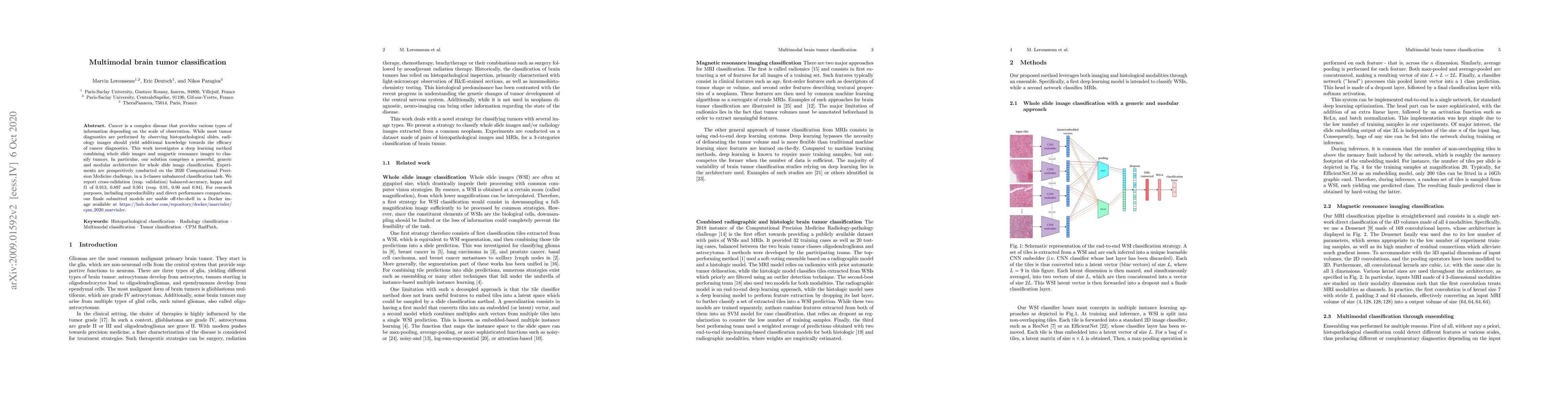
AI Quick Summary
Researchers developed a deep learning method to classify brain tumors using whole slide images and magnetic resonance images, achieving high accuracy rates in a 3-classes unbalanced classification task.
Paper Preview
Abstract
Cancer is a complex disease that provides various types of information depending on the scale of observation. While most tumor diagnostics are performed by observing histopathological slides, radiology images should yield additional knowledge towards the efficacy of cancer diagnostics. This work investigates a deep learning method combining whole slide images and magnetic resonance images to classify tumors. In particular, our solution comprises a powerful, generic and modular architecture for whole slide image classification. Experiments are prospectively conducted on the 2020 Computational Precision Medicine challenge, in a 3-classes unbalanced classification task. We report cross-validation (resp. validation) balanced-accuracy, kappa and f1 of 0.913, 0.897 and 0.951 (resp. 0.91, 0.90 and 0.94). For research purposes, including reproducibility and direct performance comparisons, our finale submitted models are usable off-the-shelf in a Docker image available at https://hub.docker.com/repository/docker/marvinler/cpm_2020_marvinler.
AI Key Findings
Get AI-generated insights about this paper's methodology, results, significance, and more — seven facets brought into focus.
Impact
Paper Details
Authors
PDF Preview
Key Terms
Citation Network
Current paper (gray), citations (green), references (blue)
Display is limited for performance on very large graphs.
Discussion 0