Multimodal Brain Tumour Classification Using Feature Fusion
Publication
Metrics
Paper Preview
Abstract
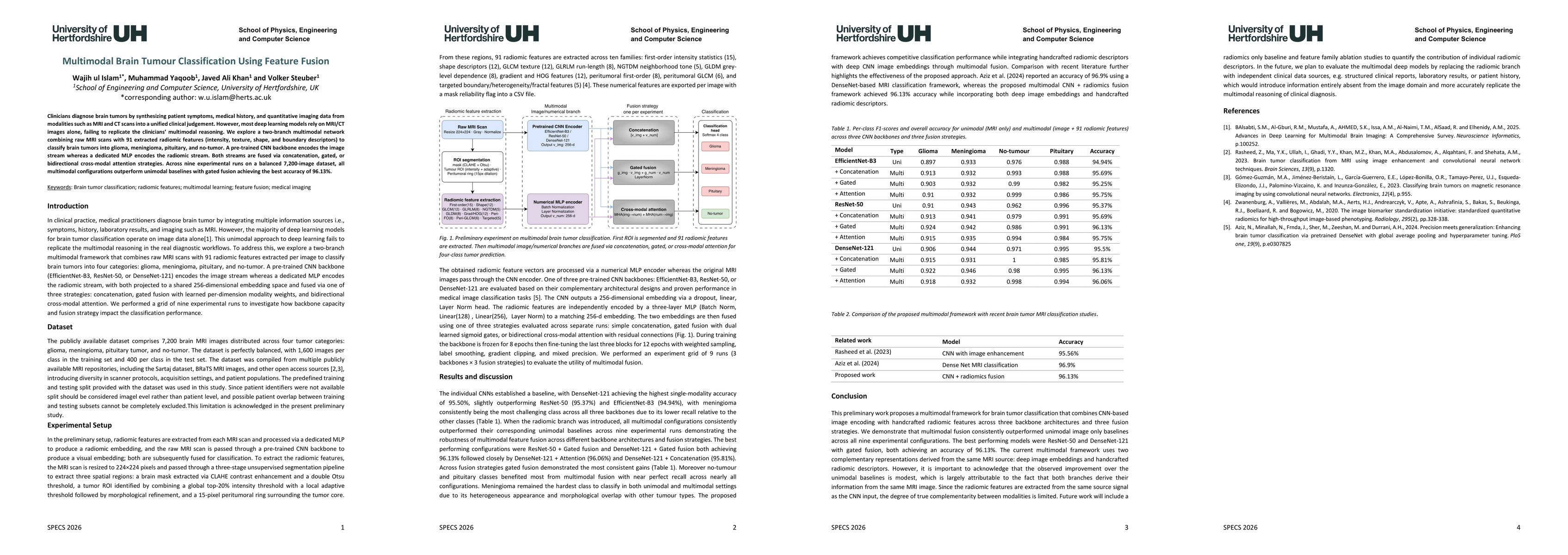
Clinicians diagnose brain tumors by synthesizing patient symptoms, medical history, and quantitative imaging data from modalities such as MRI and CT scans into a unified clinical judgement. However, most deep learning models rely on MRI/CT images alone, failing to replicate the clinicians multimodal reasoning. We explore a two-branch multimodal network combining raw MRI scans with 91 extracted radiomic features (intensity, texture, shape, and boundary descriptors) to classify brain tumors into glioma, meningioma, pituitary, and no-tumor. A pre-trained CNN backbone encodes the image stream, whereas a dedicated MLP encodes the radiomic stream. Both streams are fused via concatenation, gated, or bidirectional cross-modal attention strategies. Across nine experimental runs on a balanced 7,200 image dataset, all multimodal configurations outperform unimodal baselines with gated fusion achieving the best accuracy of 96.13%.
AI Key Findings
Get AI-generated insights about this paper's methodology, results, significance, and more — seven facets brought into focus.
Discussion 0