Authors
Publication
Metrics
Quick Actions
Quick Answers
What methodology did the authors use?
The study develops a multimodal MRI report‑supervised segmentation framework (MS‑RSuper) that parses radiology reports into quantitative global cues and qualitative modality‑specific cues using a domain‑specific LLM, then aligns these cues to substructure probability maps and trains a 3D nnU‑Net with a unified one‑sided, uncertainty‑aware loss comprising existence/absence, size/count, and cohort‑specific anatomical priors. More in Methodology →
What are the key results?
MS‑RSuper outperforms both a masks‑only baseline and a naive RSuper method on the combined BraTS‑MEN and BraTS‑MET datasets, achieving Dice scores of 0.554 (MEN WT) and 0.529 (MET WT) versus 0.452 and 0.443 for naive RSuper. — Ablation studies show that each loss component contributes: existence/absence loss gives the largest boost, global qualitative alignment further improves performance, and cohort priors... More in Key Results →
Why is this work significant?
By leveraging large‑scale radiology reports, the method reduces the need for dense voxel‑level annotations while improving multimodal brain tumor segmentation, thereby enhancing clinical decision support and accelerating deployment of AI tools in neuro‑oncology. More in Significance →
What are the main limitations?
Performance depends on accurate LLM parsing of reports; parsing errors can degrade segmentation quality. — The approach has only been validated on meningioma and metastasis cohorts; generalization to other tumor types or imaging modalities remains untested. More in Limitations →
Abstract
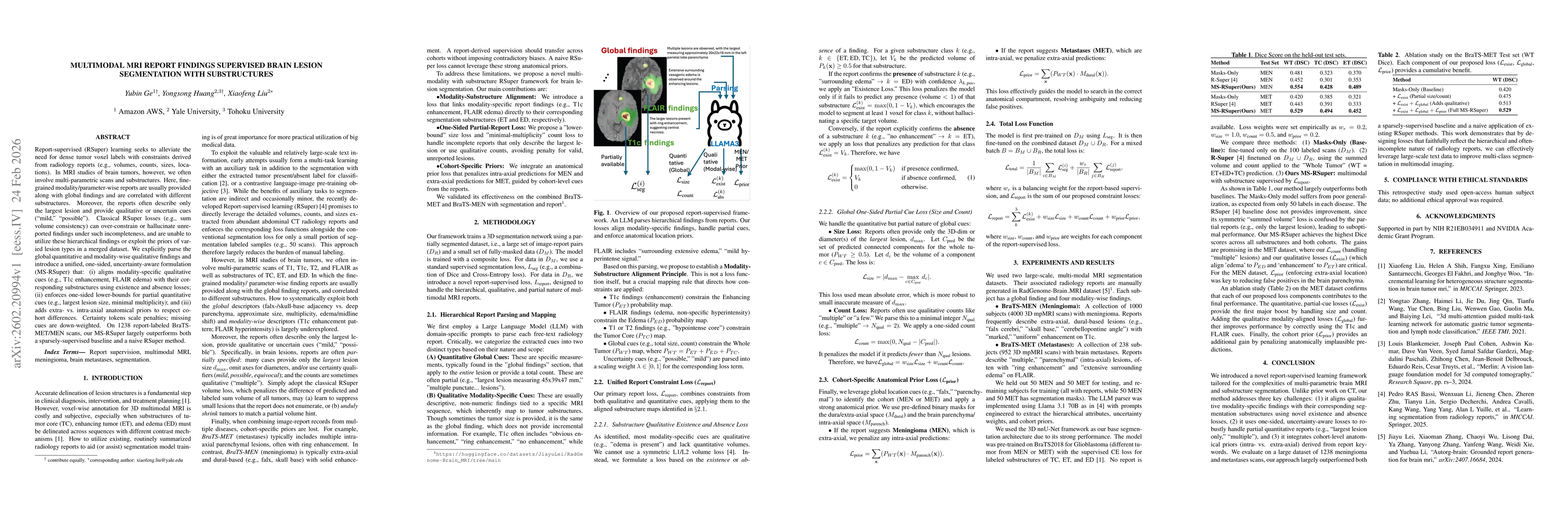
Report-supervised (RSuper) learning seeks to alleviate the need for dense tumor voxel labels with constraints derived from radiology reports (e.g., volumes, counts, sizes, locations). In MRI studies of brain tumors, however, we often involve multi-parametric scans and substructures. Here, fine-grained modality/parameter-wise reports are usually provided along with global findings and are correlated with different substructures. Moreover, the reports often describe only the largest lesion and provide qualitative or uncertain cues (``mild,'' ``possible''). Classical RSuper losses (e.g., sum volume consistency) can over-constrain or hallucinate unreported findings under such incompleteness, and are unable to utilize these hierarchical findings or exploit the priors of varied lesion types in a merged dataset. We explicitly parse the global quantitative and modality-wise qualitative findings and introduce a unified, one-sided, uncertainty-aware formulation (MS-RSuper) that: (i) aligns modality-specific qualitative cues (e.g., T1c enhancement, FLAIR edema) with their corresponding substructures using existence and absence losses; (ii) enforces one-sided lower-bounds for partial quantitative cues (e.g., largest lesion size, minimal multiplicity); and (iii) adds extra- vs. intra-axial anatomical priors to respect cohort differences. Certainty tokens scale penalties; missing cues are down-weighted. On 1238 report-labeled BraTS-MET/MEN scans, our MS-RSuper largely outperforms both a sparsely-supervised baseline and a naive RSuper method.
AI Key Findings
Generated Feb 25, 2026
Methodology — What approach did the authors take?
The study develops a multimodal MRI report‑supervised segmentation framework (MS‑RSuper) that parses radiology reports into quantitative global cues and qualitative modality‑specific cues using a domain‑specific LLM, then aligns these cues to substructure probability maps and trains a 3D nnU‑Net with a unified one‑sided, uncertainty‑aware loss comprising existence/absence, size/count, and cohort‑specific anatomical priors.
Key Results — What are the main findings?
- MS‑RSuper outperforms both a masks‑only baseline and a naive RSuper method on the combined BraTS‑MEN and BraTS‑MET datasets, achieving Dice scores of 0.554 (MEN WT) and 0.529 (MET WT) versus 0.452 and 0.443 for naive RSuper.
- Ablation studies show that each loss component contributes: existence/absence loss gives the largest boost, global qualitative alignment further improves performance, and cohort priors add additional gains.
- The framework effectively handles partial and uncertain reports, correctly aligning modality‑specific findings (e.g., T1c enhancement to enhancing tumor) to substructures and reducing false positives in anatomically implausible regions.
Significance — Why does this research matter?
By leveraging large‑scale radiology reports, the method reduces the need for dense voxel‑level annotations while improving multimodal brain tumor segmentation, thereby enhancing clinical decision support and accelerating deployment of AI tools in neuro‑oncology.
Technical Contribution — What is the technical contribution?
A unified, one‑sided, uncertainty‑aware report‑supervised loss that integrates existence/absence constraints, partial quantitative bounds, and cohort‑specific anatomical priors, enabling effective alignment of modality‑specific report cues to substructure segmentation.
Novelty — What is new about this work?
First to address multimodal MRI substructure segmentation with hierarchical report parsing, handling partial/uncertain reports via one‑sided losses, and incorporating cohort‑specific anatomical priors—features absent in prior RSuper work.
Limitations — What are the limitations of this study?
- Performance depends on accurate LLM parsing of reports; parsing errors can degrade segmentation quality.
- The approach has only been validated on meningioma and metastasis cohorts; generalization to other tumor types or imaging modalities remains untested.
Future Work — What did the authors propose for future work?
- Extend the framework to additional brain tumor types and multi‑disease cohorts, incorporating more diverse reports.
- Improve LLM parsing accuracy and uncertainty modeling, possibly by joint end‑to‑end training of the language model and segmentation network.
Paper Details
How to Cite This Paper
@article{liu2026multimodal,
title = {Multimodal MRI Report Findings Supervised Brain Lesion Segmentation with Substructures},
author = {Liu, Xiaofeng and Huang, Yongsong and Ge, Yubin},
year = {2026},
eprint = {2602.20994},
archivePrefix = {arXiv},
primaryClass = {eess.IV},
}
Liu, X., Huang, Y., & Ge, Y. (2026). Multimodal MRI Report Findings Supervised Brain Lesion Segmentation with Substructures. arXiv. https://arxiv.org/abs/2602.20994
Liu, Xiaofeng, et al. "Multimodal MRI Report Findings Supervised Brain Lesion Segmentation with Substructures." arXiv, 2026, arxiv.org/abs/2602.20994.
PDF Preview
Similar Papers
Found 4 papersAutomatic Rodent Brain MRI Lesion Segmentation with Fully Convolutional Networks
Jussi Tohka, Riccardo de Feo, Artem Shatillo et al.
Interpretable Brain MRI Report Generation Anchored by Lesion Topography.
Zhang, Xiaoman, Li, Yuehua, Zhang, Ya et al.
Combining CNNs With Transformer for Multimodal 3D MRI Brain Tumor Segmentation With Self-Supervised Pretraining
Ostap Viniavskyi, Mariia Dobko, Danylo-Ivan Kolinko et al.
Comments (0)