Multiple sclerosis lesion enhancement and white matter region estimation using hyperintensities in FLAIR images
Publication
Metrics
AI Quick Summary
This paper proposes a lesion enhancement technique for FLAIR MRI images to better distinguish multiple sclerosis lesions from white and gray matter. The method significantly improves lesion brightness compared to surrounding tissues, aiding both expert diagnosis and automated segmentation, and also enables better estimation of white matter regions.
Paper Preview
Abstract
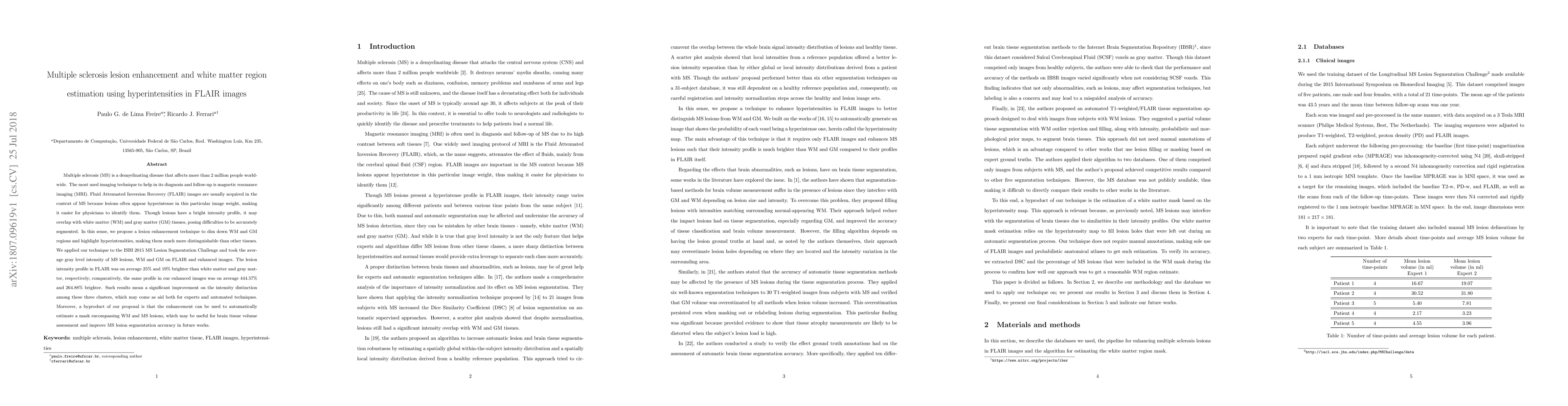
Multiple sclerosis (MS) is a demyelinating disease that affects more than 2 million people worldwide. The most used imaging technique to help in its diagnosis and follow-up is magnetic resonance imaging (MRI). Fluid Attenuated Inversion Recovery (FLAIR) images are usually acquired in the context of MS because lesions often appear hyperintense in this particular image weight, making it easier for physicians to identify them. Though lesions have a bright intensity profile, it may overlap with white matter (WM) and gray matter (GM) tissues, posing difficulties to be accurately segmented. In this sense, we propose a lesion enhancement technique to dim down WM and GM regions and highlight hyperintensities, making them much more distinguishable than other tissues. We applied our technique to the ISBI 2015 MS Lesion Segmentation Challenge and took the average gray level intensity of MS lesions, WM and GM on FLAIR and enhanced images. The lesion intensity profile in FLAIR was on average 25% and 19% brighter than white matter and gray matter, respectively; comparatively, the same profile in our enhanced images was on average 444% and 264% brighter. Such results mean a significant improvement on the intensity distinction among these three clusters, which may come as aid both for experts and automated techniques. Moreover, a byproduct of our proposal is that the enhancement can be used to automatically estimate a mask encompassing WM and MS lesions, which may be useful for brain tissue volume assessment and improve MS lesion segmentation accuracy in future works.
AI Key Findings
Get AI-generated insights about this paper's methodology, results, significance, and more — seven facets brought into focus.
Impact
Paper Details
PDF Preview
Key Terms
Citation Network
Current paper (gray), citations (green), references (blue)
Display is limited for performance on very large graphs.
Discussion 0