Publication
Metrics
AI Quick Summary
This paper proposes a method to enhance spectral domain optical coherence tomography by using phase modulators and delay lines to increase axial resolution and maximum unambiguous range beyond the Nyquist limit. Simulation results demonstrate that combining multiple channels via digital filter banks can significantly improve system performance.
Paper Preview
Abstract
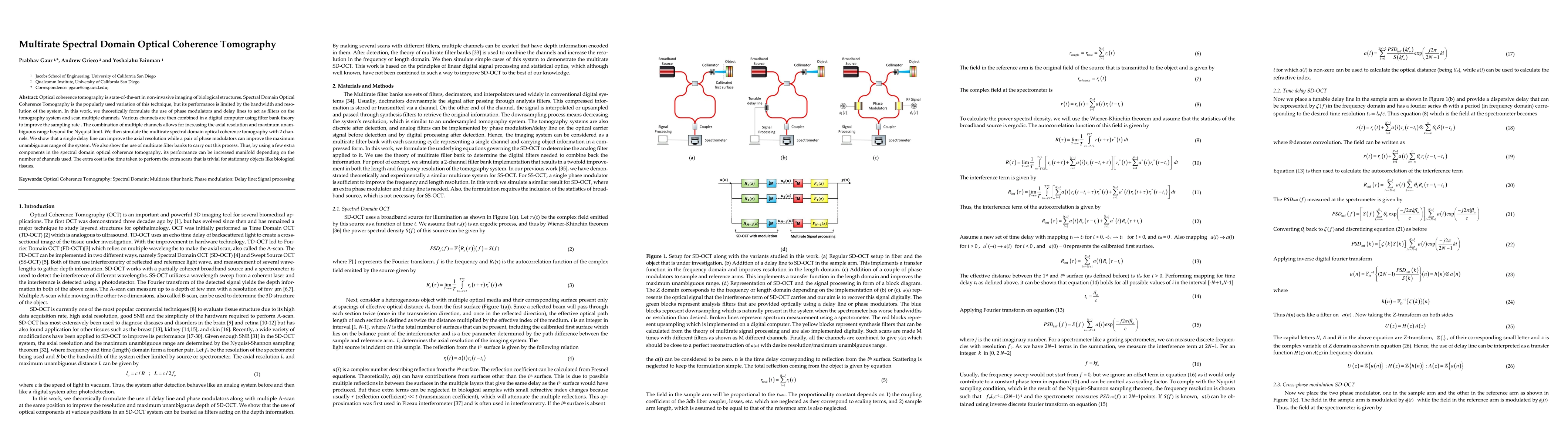
Optical coherence tomography is state-of-the-art in non-invasive imaging of biological structures. Spectral Domain Optical Co-herence Tomography is the popularly used variation of this technique, but its performance is limited by the bandwidth and res-olution of the system. In this work, we theoretically formulate the use of phase modulators and delay lines to act as filters on the tomography system and scan multiple channels. Various channels are then combined in a digital computer using filter bank theory to improve the sampling rate . The combination of multiple channels allows for increasing the axial resolution and maximum unambiguous range beyond the Nyquist limit. We then simulate the multirate spectral domain optical coherence tomography with 2 channels. We show that a single delay line can improve the axial resolution while a pair of phase modulators can improve the maximum unambiguous range of the system. We also show the use of multirate filter banks to carry out this process. Thus, by using a few extra components in the spectral domain optical coherence tomography, its performance can be increased manifold de-pending on the number of channels used. The extra cost is the time taken to perform the extra scans that is trivial for stationary objects like biological tissues.
AI Key Findings
Get AI-generated insights about this paper's methodology, results, significance, and more — seven facets brought into focus.
Impact
Paper Details
Authors
PDF Preview
Key Terms
Citation Network
Current paper (gray), citations (green), references (blue)
Display is limited for performance on very large graphs.
Discussion 0