Publication
Metrics
AI Quick Summary
This research investigates structural differences between mouse and human neurons and applies these insights to generative AIs. Mouse neurons, with their smaller size and thinner neurites, were implemented in AI models, which outperformed standard models in generating images of cats and cheese but underperformed for human faces and birds, suggesting dataset-specific preferences.
Paper Preview
Abstract
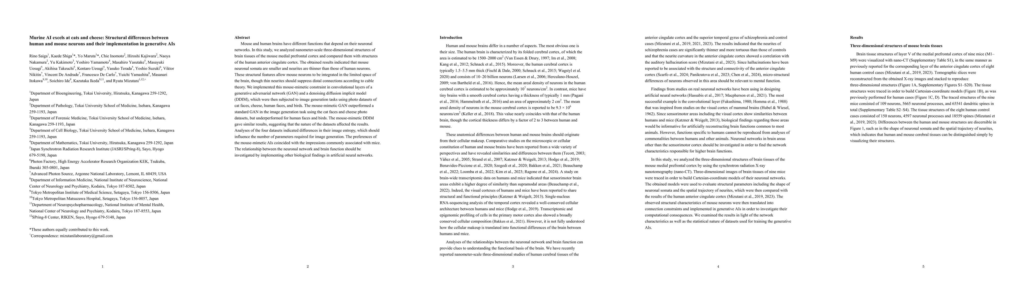
Mouse and human brains have different functions that depend on their neuronal networks. In this study, we analyzed nanometer-scale three-dimensional structures of brain tissues of the mouse medial prefrontal cortex and compared them with structures of the human anterior cingulate cortex. The obtained results indicated that mouse neuronal somata are smaller and neurites are thinner than those of human neurons. These structural features allow mouse neurons to be integrated in the limited space of the brain, though thin neurites should suppress distal connections according to cable theory. We implemented this mouse-mimetic constraint in convolutional layers of a generative adversarial network (GAN) and a denoising diffusion implicit model (DDIM), which were then subjected to image generation tasks using photo datasets of cat faces, cheese, human faces, and birds. The mouse-mimetic GAN outperformed a standard GAN in the image generation task using the cat faces and cheese photo datasets, but underperformed for human faces and birds. The mouse-mimetic DDIM gave similar results, suggesting that the nature of the datasets affected the results. Analyses of the four datasets indicated differences in their image entropy, which should influence the number of parameters required for image generation. The preferences of the mouse-mimetic AIs coincided with the impressions commonly associated with mice. The relationship between the neuronal network and brain function should be investigated by implementing other biological findings in artificial neural networks.
AI Key Findings
Get AI-generated insights about this paper's methodology, results, significance, and more — seven facets brought into focus.
Impact
Paper Details
Authors
PDF Preview
Citation Network
Current paper (gray), citations (green), references (blue)
Display is limited for performance on very large graphs.
Discussion 0