Nanoparticle-assisted STED nanoscopy with gold nanospheres
Publication
Metrics
AI Quick Summary
Researchers developed a new method for STED nanoscopy using gold nanospheres coated with fluorescent silica, achieving better resolution and brighter images than traditional methods, with potential applications in bio-imaging.
Paper Preview
Abstract
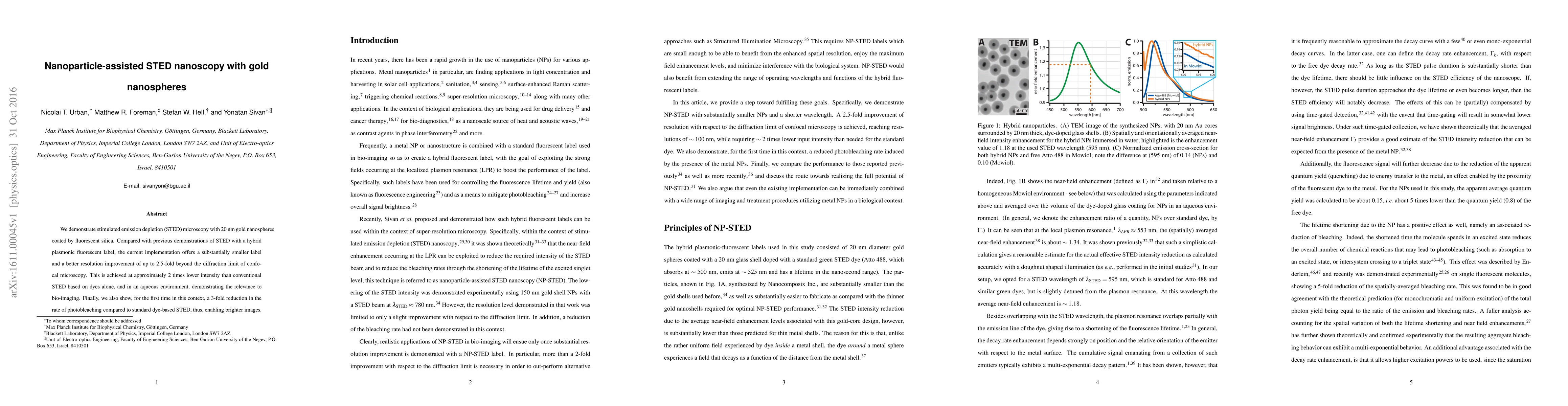
We demonstrate stimulated emission depletion (STED) microscopy with 20 nm gold nanospheres coated by fluorescent silica. Compared with previous demonstrations of STED with a hybrid plasmonic fluorescent label, the current implementation offers a substantially smaller label and a better resolution improvement of up to 2.5-fold beyond the diffraction limit of confocal microscopy. This is achieved at approximately 2 times lower intensity than conventional STED based on dyes alone, and in an aqueous environment, demonstrating the relevance to bio-imaging. Finally, we also show, for the first time in this context, a 3-fold reduction in the rate of photobleaching compared to standard dye-based STED, thus, enabling brighter images.
AI Key Findings
Get AI-generated insights about this paper's methodology, results, significance, and more — seven facets brought into focus.
Impact
Paper Details
PDF Preview
Key Terms
Citation Network
Current paper (gray), citations (green), references (blue)
Display is limited for performance on very large graphs.
Discussion 0