Nanoparticle-Protein Interaction: Demystifying the Correlation Between Protein Corona and Aggregation Phenomena
Publication
Metrics
AI Quick Summary
This study employs a multi-technique approach to distinguish protein corona from nanoparticle aggregation, using Dynamic Light Scattering, Small-Angle X-ray Scattering, and cryo-TEM. The methodology successfully differentiates between protein corona, small aggregates, and massive aggregation in model silica nanoparticles incubated with various proteins.
Paper Preview
Abstract
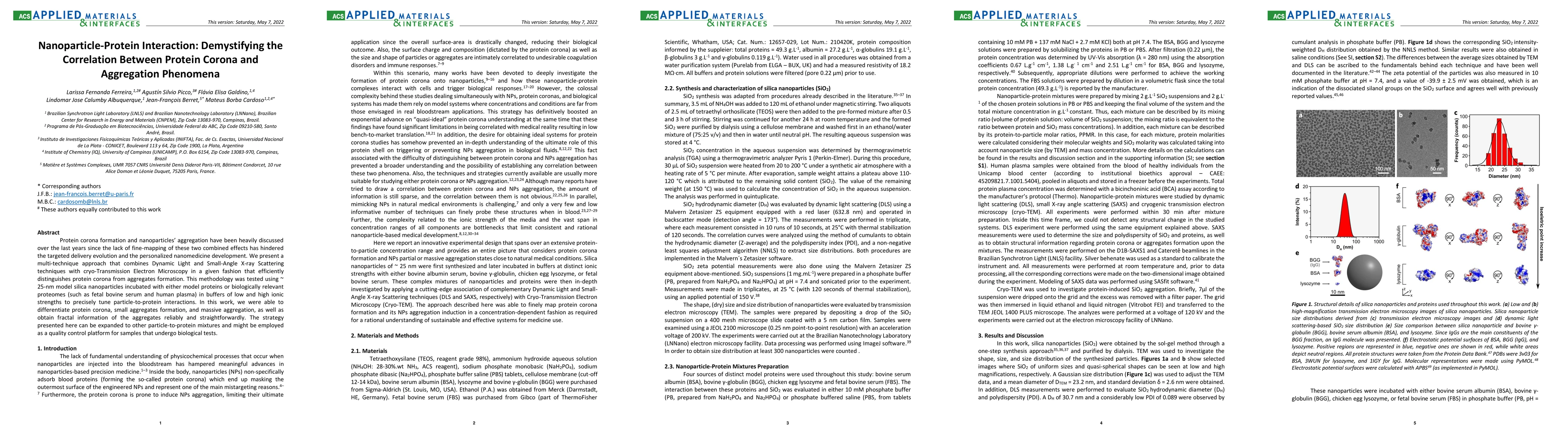
Protein corona formation and nanoparticle aggregation have been heavily discussed over the last years since the lack of fine-mapping of these two combined effects has hindered the targeted delivery evolution and the personalized nanomedicine development. We present a multi-technique approach that combines Dynamic Light and Small-Angle X-ray Scattering techniques with cryo-Transmission Electron Microscopy in a given fashion that efficiently distinguishes protein corona from aggregates formation. This methodology was tested using 25-nm model silica nanoparticles incubated with either model proteins or biologically relevant proteomes (such as fetal bovine serum and human plasma) in buffers of low and high ionic strengths to precisely tune particle-to-protein interactions. In this work, we were able to differentiate protein corona, small aggregates formation, and massive aggregation, as well as obtain fractal information of the aggregates reliably and straightforwardly. The strategy presented here can be expanded to other particle-to-protein mixtures and might be employed as a quality control platform for samples that undergo biological tests.
AI Key Findings
Get AI-generated insights about this paper's methodology, results, significance, and more — seven facets brought into focus.
Impact
Paper Details
Authors
PDF Preview
Key Terms
Citation Network
Current paper (gray), citations (green), references (blue)
Display is limited for performance on very large graphs.
Discussion 0