Morphometry of medial temporal lobe (MTL) subregions in brain MRI is
sensitive biomarker to Alzheimers Disease and other related conditions. While
T2-weighted (T2w) MRI with high in-plane resolution is widely used to segment
hippocampal subfields due to its higher contrast in hippocampus, its lower
out-of-plane resolution reduces the accuracy of subregion thickness
measurements. To address this issue, we developed a nearly isotropic
segmentation pipeline that incorporates image and label upsampling and
high-resolution segmentation in T2w MRI. First, a high-resolution atlas was
created based on an existing anisotropic atlas derived from 29 individuals.
Both T1-weighted and T2w images in the atlas were upsampled from their original
resolution to a nearly isotropic resolution 0.4x0.4x0.52mm3 using a non-local
means approach. Manual segmentations within the atlas were also upsampled to
match this resolution using a UNet-based neural network, which was trained on a
cohort consisting of both high-resolution ex vivo and low-resolution
anisotropic in vivo MRI with manual segmentations. Second, a multi-modality
deep learning-based segmentation model was trained within this nearly isotropic
atlas. Finally, experiments showed the nearly isotropic subregion segmentation
improved the accuracy of cortical thickness as an imaging biomarker for
neurodegeneration in T2w MRI.
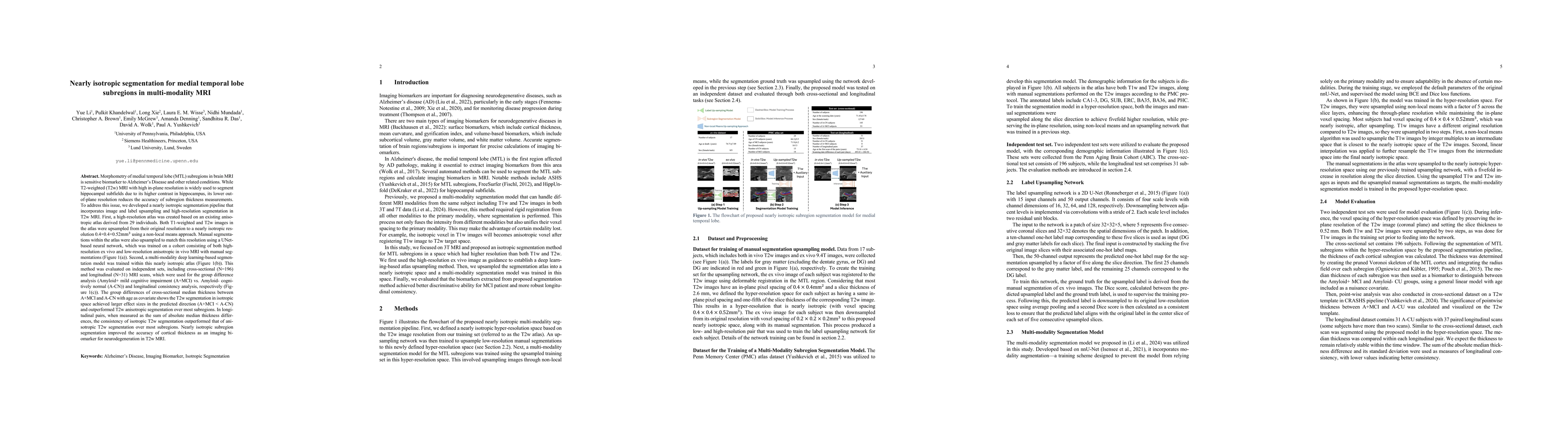
Discussion 0