Network analysis on cortical morphometry in first-episode schizophrenia
Publication
Metrics
AI Quick Summary
This study investigates cortical morphological connectivity in first-episode schizophrenia (FES) using network analysis on cortical thickness and surface area from MRI scans. Results show that FES patients exhibit reduced local connectivity and small-worldness in cortical networks, particularly in frontal, parietal, and temporal lobes, suggesting potential as a biomarker for FES.
Paper Preview
Abstract
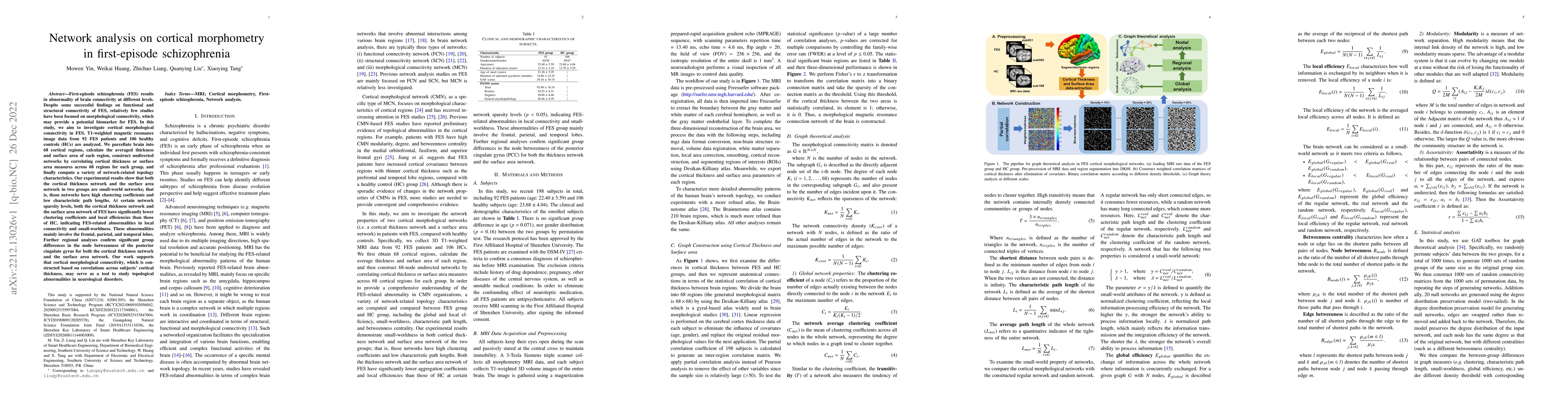
First-episode schizophrenia (FES) results in abnormality of brain connectivity at different levels. Despite some successful findings on functional and structural connectivity of FES, relatively few studies have been focused on morphological connectivity, which may provide a potential biomarker for FES. In this study, we aim to investigate cortical morphological connectivity in FES. T1-weighted magnetic resonance image data from 92 FES patients and 106 healthy controls (HCs) are analyzed.We parcellate brain into 68 cortical regions, calculate the averaged thickness and surface area of each region, construct undirected networks by correlating cortical thickness or surface area measures across 68 regions for each group, and finally compute a variety of network-related topology characteristics. Our experimental results show that both the cortical thickness network and the surface area network in two groups are small-world networks; that is, those networks have high clustering coefficients and low characteristic path lengths. At certain network sparsity levels, both the cortical thickness network and the surface area network of FES have significantly lower clustering coefficients and local efficiencies than those of HC, indicating FES-related abnormalities in local connectivity and small-worldness. These abnormalities mainly involve the frontal, parietal, and temporal lobes. Further regional analyses confirm significant group differences in the node betweenness of the posterior cingulate gyrus for both the cortical thickness network and the surface area network. Our work supports that cortical morphological connectivity, which is constructed based on correlations across subjects' cortical thickness, may serve as a tool to study topological abnormalities in neurological disorders.
AI Key Findings
Get AI-generated insights about this paper's methodology, results, significance, and more — seven facets brought into focus.
Impact
Paper Details
Authors
PDF Preview
Key Terms
Citation Network
Current paper (gray), citations (green), references (blue)
Display is limited for performance on very large graphs.
Discussion 0