Neurodevelopmental Age Estimation of Infants Using a 3D-Convolutional Neural Network Model based on Fusion MRI Sequences
Publication
Metrics
AI Quick Summary
This study developed a 3D convolutional neural network model to estimate brain developmental age in infants using fusion MRI sequences, achieving high precision and recall, which could aid in identifying developmental delays and neurological diseases. The method uses T1, T2, and PD MRI sequences to provide an objective measure of brain maturity.
Paper Preview
Abstract
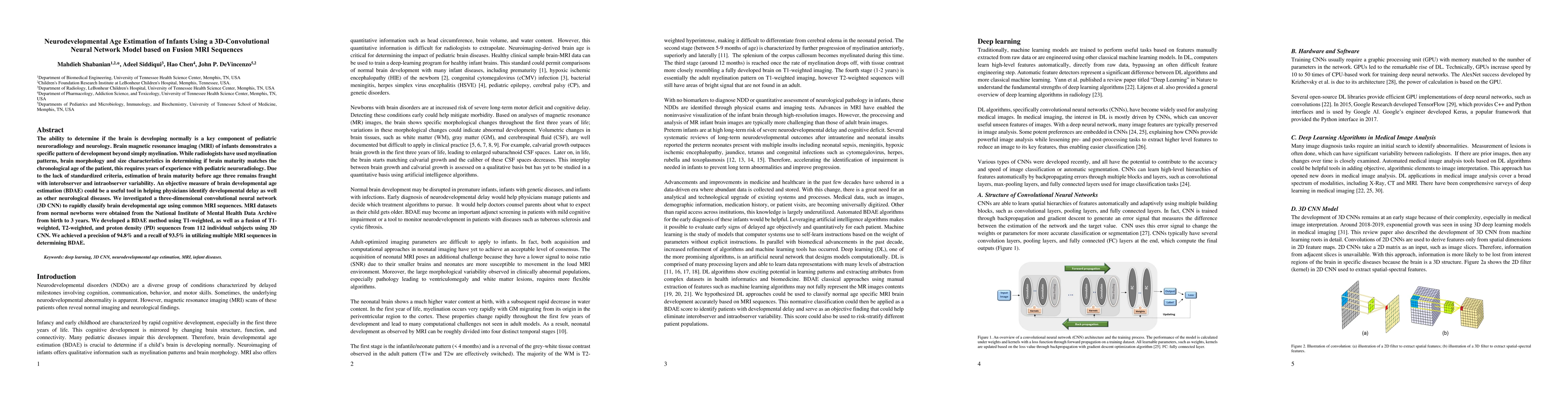
The ability to determine if the brain is developing normally is a key component of pediatric neuroradiology and neurology. Brain magnetic resonance imaging (MRI) of infants demonstrates a specific pattern of development beyond simply myelination. While radiologists have used myelination patterns, brain morphology and size characteristics in determining if brain maturity matches the chronological age of the patient, this requires years of experience with pediatric neuroradiology. Due to the lack of standardized criteria, estimation of brain maturity before age three remains fraught with interobserver and intraobserver variability. An objective measure of brain developmental age estimation (BDAE) could be a useful tool in helping physicians identify developmental delay as well as other neurological diseases. We investigated a three-dimensional convolutional neural network (3D CNN) to rapidly classify brain developmental age using common MRI sequences. MRI datasets from normal newborns were obtained from the National Institute of Mental Health Data Archive from birth to 3 years. We developed a BDAE method using T1-weighted, as well as a fusion of T1-weighted, T2-weighted, and proton density (PD) sequences from 112 individual subjects using 3D CNN. We achieved a precision of 94.8% and a recall of 93.5% in utilizing multiple MRI sequences in determining BDAE.
AI Key Findings
Get AI-generated insights about this paper's methodology, results, significance, and more — seven facets brought into focus.
Impact
Paper Details
Authors
PDF Preview
Key Terms
Citation Network
Current paper (gray), citations (green), references (blue)
Display is limited for performance on very large graphs.
Discussion 0