Publication
Metrics
AI Quick Summary
This paper presents innovative approaches for particle-induced prompt gamma imaging, including a knife-edge PET method for proton range verification, a fiducial marker technique for proton localization, and a novel position-sensitive gamma imaging design for 3D imaging. Simulation and experimental results demonstrate promising spatial resolution and detection efficiency for these methods.
Paper Preview
Abstract
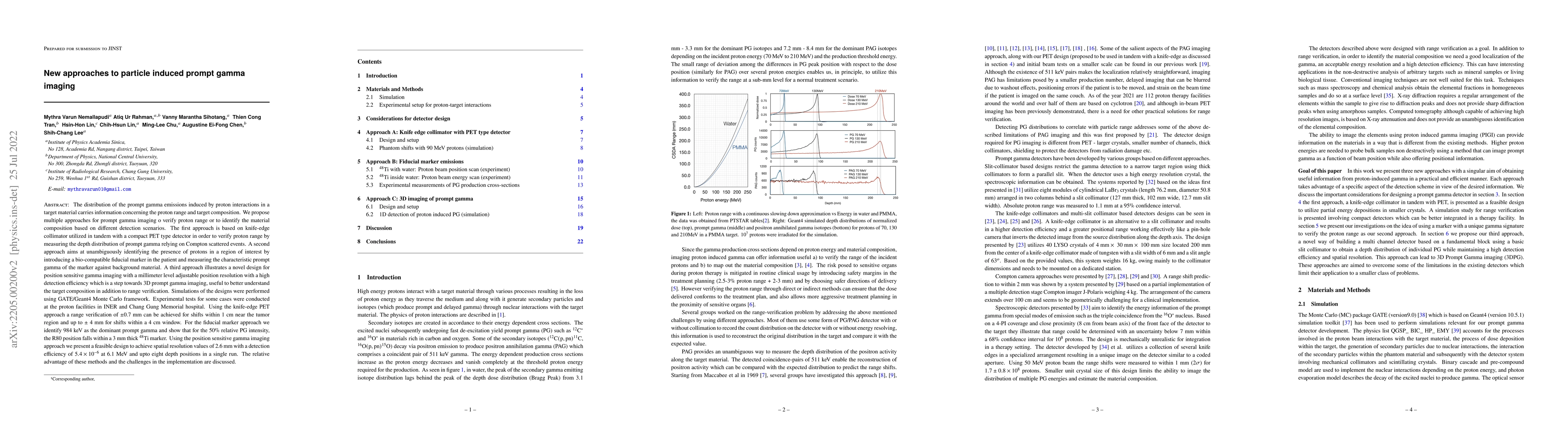
The distribution of the prompt gamma emissions induced by proton interactions in a target material carries information concerning the proton range and target composition. In order to image prompt gamma we present different approaches. The first approach is based on knife-edge collimator utilized in tandem with a compact PET type detector in order to verify proton range by measuring the depth distribution of prompt gamma relying on Compton scattered events. A second approach aims at unambiguously identifying the presence of protons in a region of interest by introducing a bio-compatible fiducial marker in the patient and measuring the characteristic prompt gamma of the marker against background material. A third approach illustrates a novel design for position sensitive gamma imaging with a millimeter level adjustable position resolution with a high detection efficiency which is a step towards 3D prompt gamma imaging, useful to better understand the target composition. Simulations of the designs were performed using GATE/Geant4 Monte Carlo framework. Experimental tests on fiducial marker were conducted at the proton facilities in INER and Chang Gung Memorial hospital. Using the knife-edge PET approach a range verification of 0.7 mm can be achieved for shifts within 1 cm near the tumor region and up to 4 mm for shifts within a 4 cm window. For the fiducial marker approach we identify 984 keV as the dominant prompt gamma and show that for the 50% relative PG intensity, the R80 position falls within a 3 mm thick 48Ti marker. Using the position sensitive gamma imaging approach we present a feasible design to achieve spatial resolution values of 2.6 mm with a detection efficiency of 5.4E-6 at 6.1 MeV and up to eight depth positions in a single run. The relative advantage of these methods and the challenges in the implementation are discussed.
AI Key Findings
Get AI-generated insights about this paper's methodology, results, significance, and more — seven facets brought into focus.
Impact
Paper Details
Authors
PDF Preview
Key Terms
Citation Network
Current paper (gray), citations (green), references (blue)
Display is limited for performance on very large graphs.
Discussion 0