Publication
Metrics
AI Quick Summary
This paper presents an experimental verification of diffusion pore imaging using only short gradient pulses, enabling flexible sequence design and faster convergence. The phase detection for pore image reconstruction is achieved via double diffusion encoding, while both magnitude and phase artifacts are analyzed.
Paper Preview
Abstract
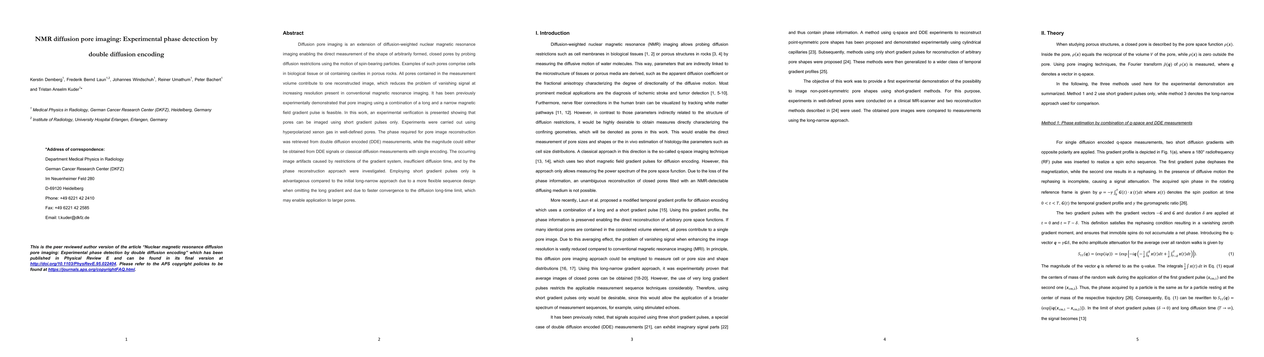
Diffusion pore imaging is an extension of diffusion-weighted nuclear magnetic resonance imaging enabling the direct measurement of the shape of arbitrarily formed, closed pores by probing diffusion restrictions using the motion of spin-bearing particles. Examples of such pores comprise cells in biological tissue or oil containing cavities in porous rocks. All pores contained in the measurement volume contribute to one reconstructed image, which reduces the problem of vanishing signal at increasing resolution present in conventional magnetic resonance imaging. It has been previously experimentally demonstrated that pore imaging using a combination of a long and a narrow magnetic field gradient pulse is feasible. In this work, an experimental verification is presented showing that pores can be imaged using short gradient pulses only. Experiments were carried out using hyperpolarized xenon gas in well-defined pores. The phase required for pore image reconstruction was retrieved from double diffusion encoded (DDE) measurements, while the magnitude could either be obtained from DDE signals or classical diffusion measurements with single encoding. The occurring image artifacts caused by restrictions of the gradient system, insufficient diffusion time, and by the phase reconstruction approach were investigated. Employing short gradient pulses only is advantageous compared to the initial long-narrow approach due to a more flexible sequence design when omitting the long gradient and due to faster convergence to the diffusion long-time limit, which may enable application to larger pores.
AI Key Findings
Get AI-generated insights about this paper's methodology, results, significance, and more — seven facets brought into focus.
Impact
Paper Details
Authors
PDF Preview
Key Terms
Citation Network
Current paper (gray), citations (green), references (blue)
Display is limited for performance on very large graphs.
Discussion 0