Noisy network attractor models for transitions between EEG microstates
Publication
Metrics
AI Quick Summary
Researchers propose a novel modeling approach for EEG microstates using noisy network attractor models to capture their dynamics and temporal correlations. These models can reproduce the transition probabilities between microstates but struggle with heavy-tailed residence time distributions.
Paper Preview
Abstract
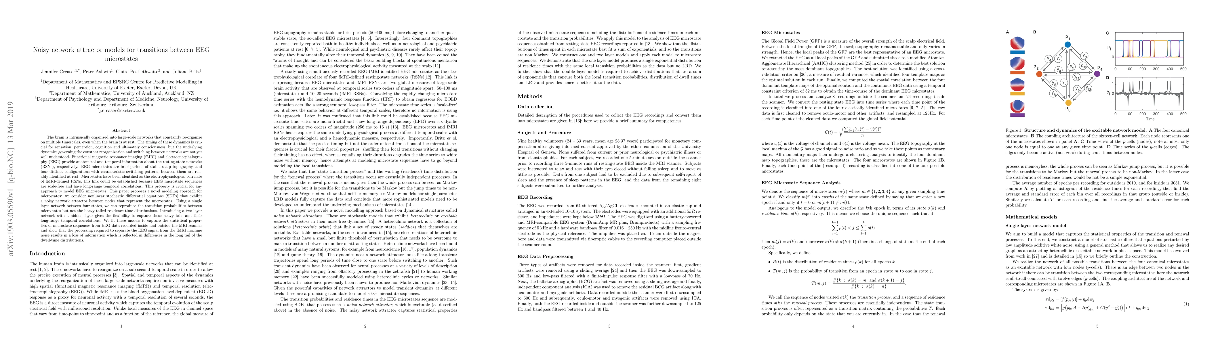
The brain is intrinsically organized into large-scale networks that constantly re-organize on multiple timescales, even when the brain is at rest. The timing of these dynamics is crucial for sensation, perception, cognition and ultimately consciousness, but the underlying dynamics governing the constant reorganization and switching between networks are not yet well understood. Functional magnetic resonance imaging (fMRI) and electroencephalography (EEG) provide anatomical and temporal information about the resting-state networks (RSNs), respectively. EEG microstates are brief periods of stable scalp topography, and four distinct configurations with characteristic switching patterns between them are reliably identified at rest. Microstates have been identified as the electrophysiological correlate of fMRI-defined RSNs, this link could be established because EEG microstate sequences are scale-free and have long-range temporal correlations. This property is crucial for any approach to model EEG microstates. This paper proposes a novel modeling approach for microstates: we consider nonlinear stochastic differential equations (SDEs) that exhibit a noisy network attractor between nodes that represent the microstates. Using a single layer network between four states, we can reproduce the transition probabilities between microstates but not the heavy tailed residence time distributions. Introducing a two layer network with a hidden layer gives the flexibility to capture these heavy tails and their long-range temporal correlations. We fit these models to capture the statistical properties of microstate sequences from EEG data recorded inside and outside the MRI scanner and show that the processing required to separate the EEG signal from the fMRI machine noise results in a loss of information which is reflected in differences in the long tail of the dwell-time distributions.
AI Key Findings
Get AI-generated insights about this paper's methodology, results, significance, and more — seven facets brought into focus.
Impact
Paper Details
PDF Preview
Key Terms
Citation Network
Current paper (gray), citations (green), references (blue)
Display is limited for performance on very large graphs.
Discussion 0